Total Pageviews

Saturday 9 June 2018

CASE 497: MULTIPLE SKIN LESIONS AND SUBCUTANEOUS TUBERCULOSIS ABSCESS , Dr PHAN THANH HẢI, MEDIC MEDICAL CENTER, HCMC, VIETNAM.


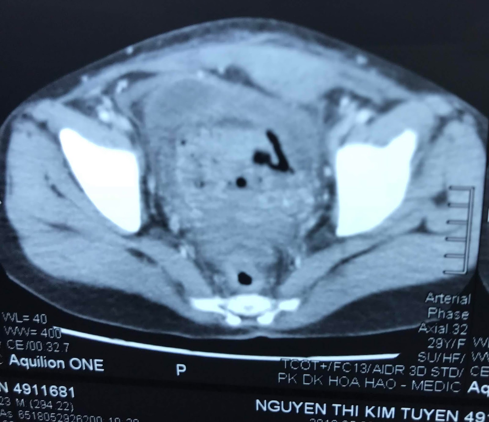
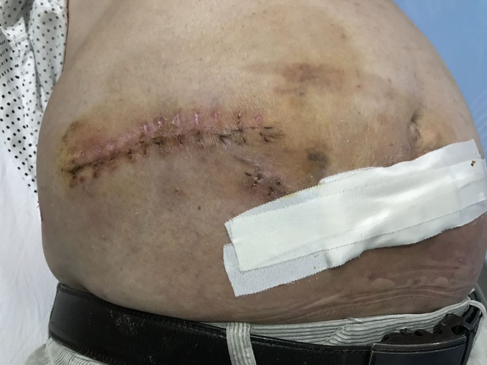
Man  47 yo  with pain at RLAQ at palpation   looked  like   appendicitis  and  skin of right leg  inflammed red and bulleous  growth  with multiple nodules.( see photo of abdomen and  leg skin).


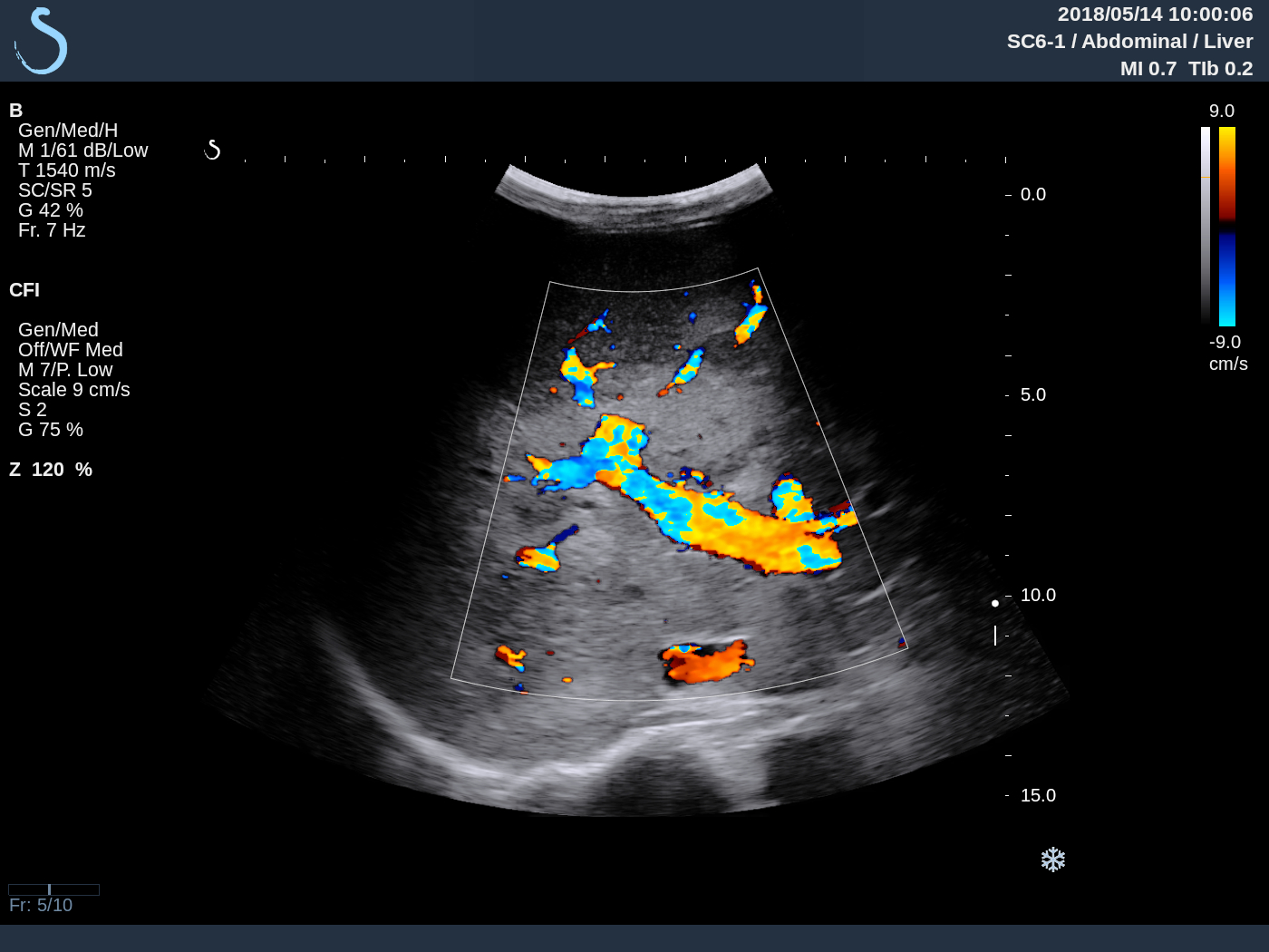
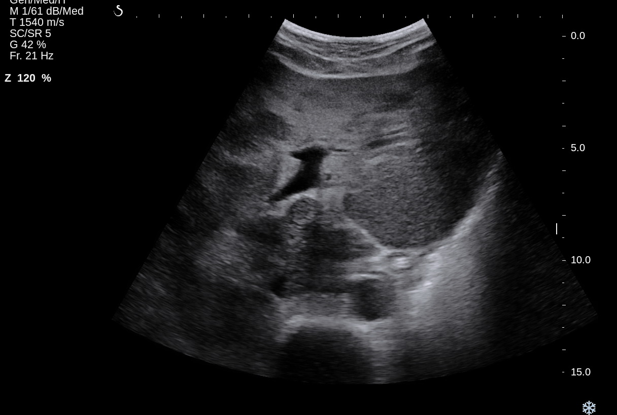
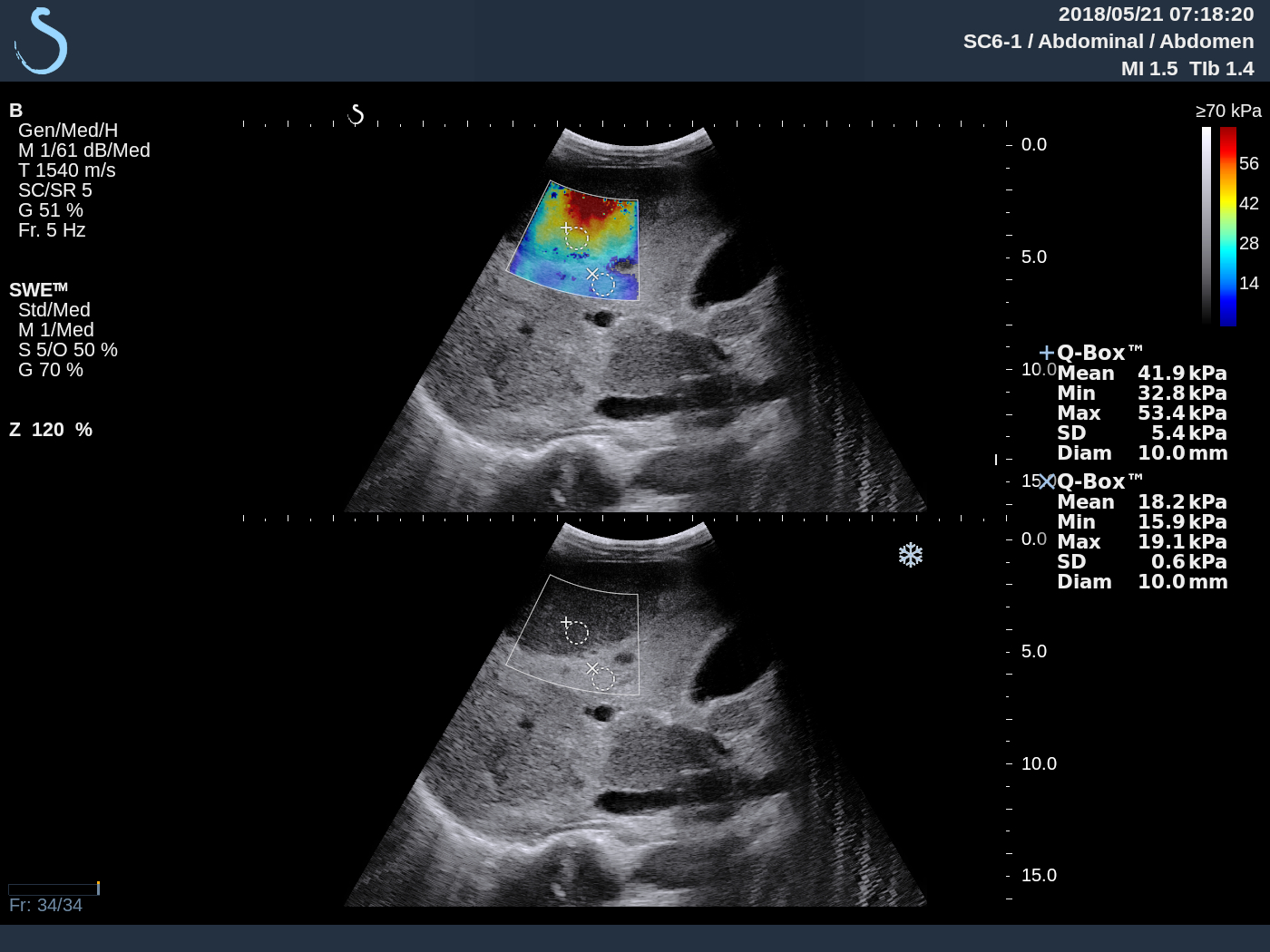
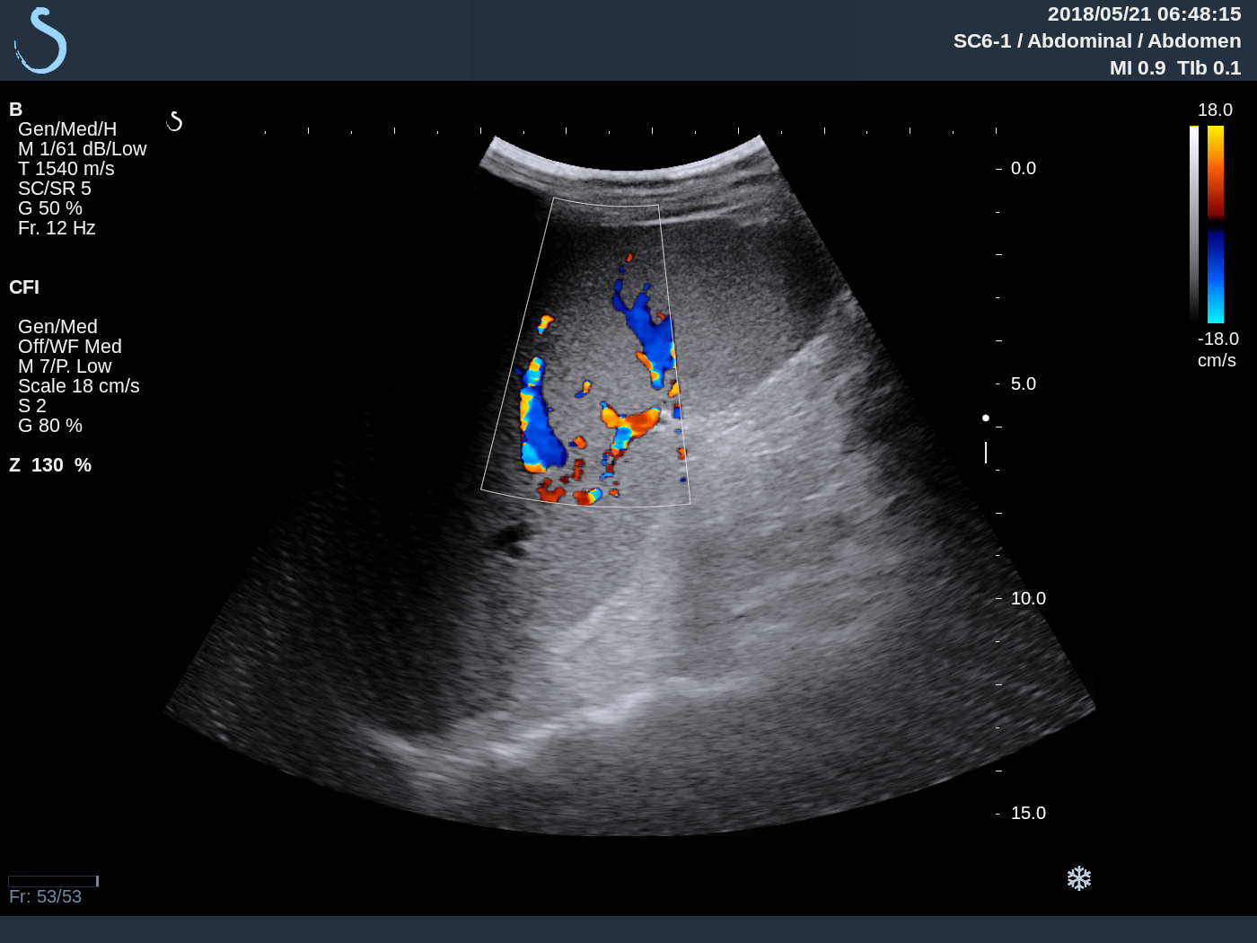
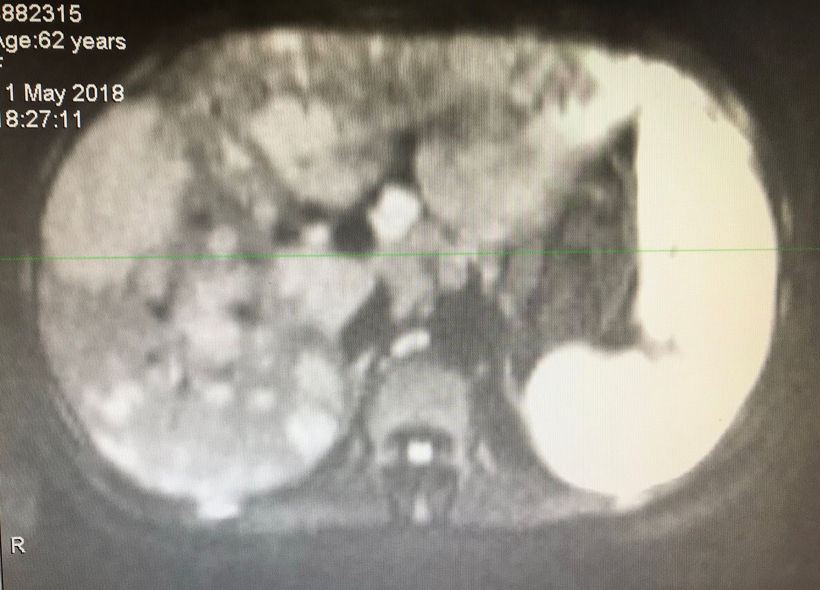
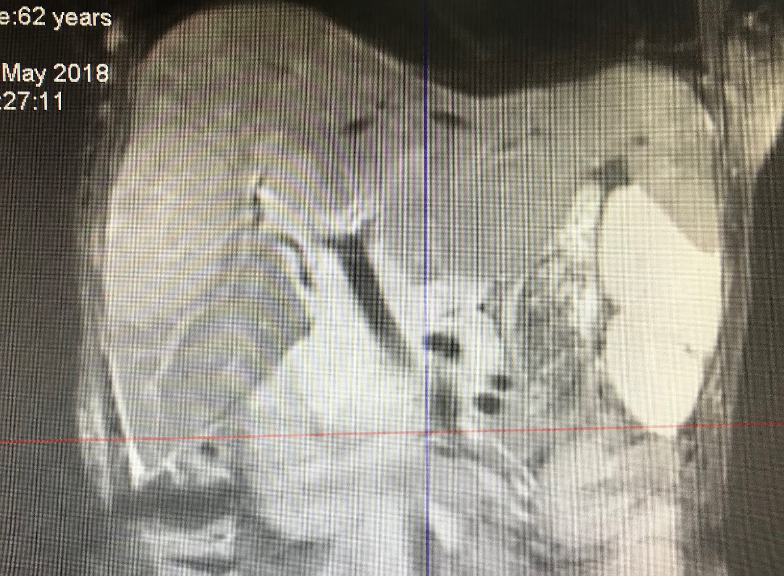
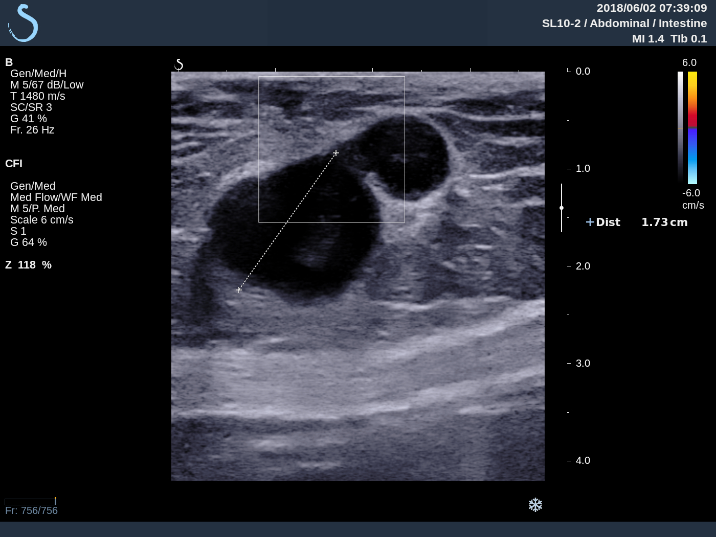
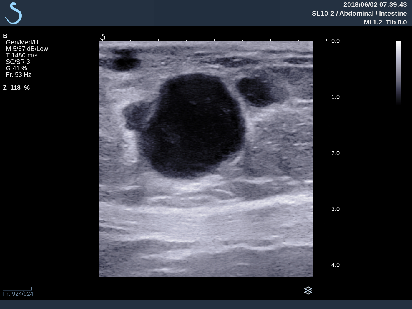
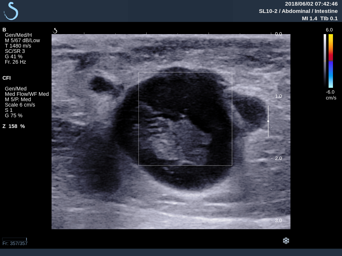
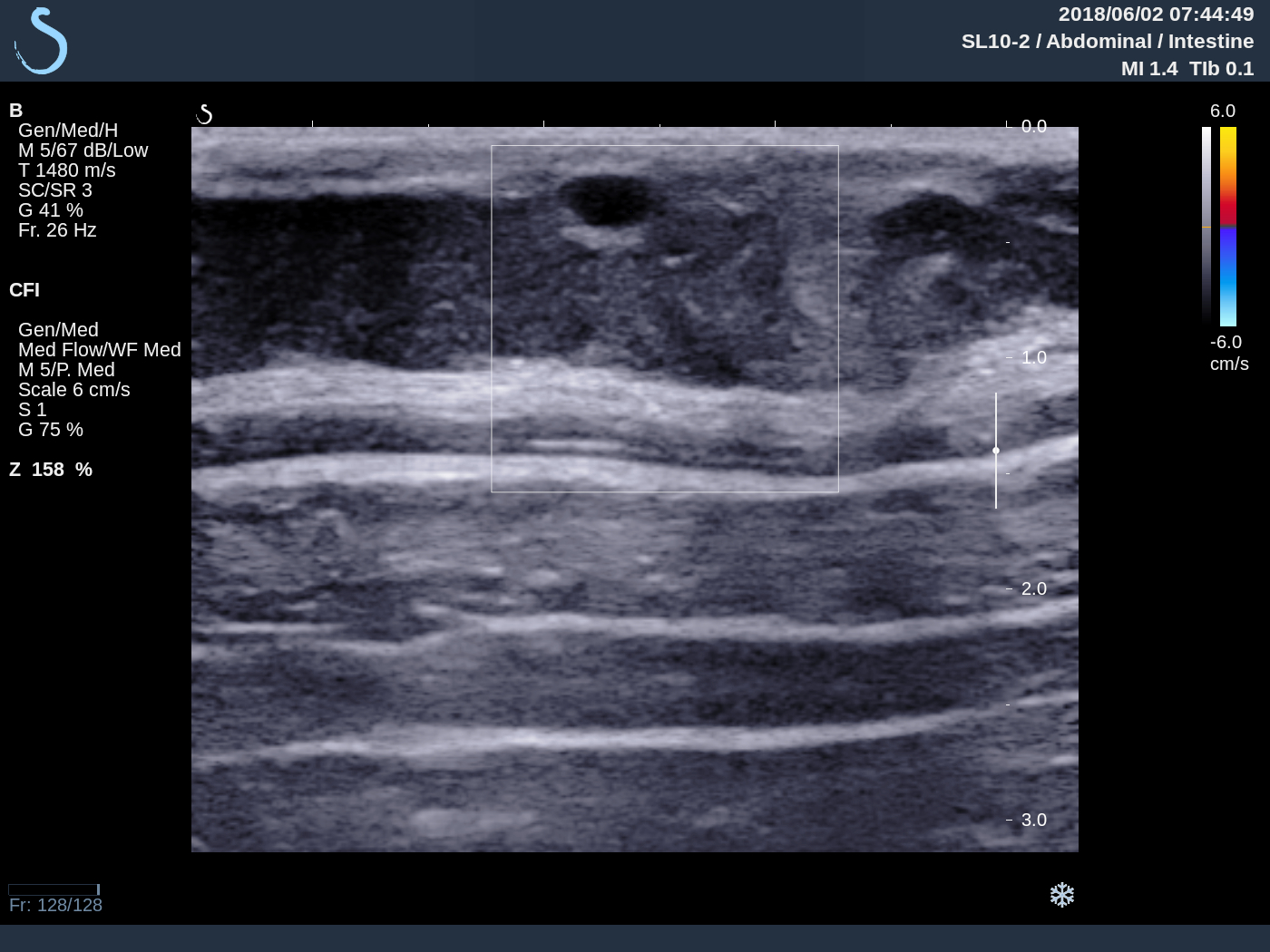
Ultrasound  of abdominal wall and the leg skin  lesions detected many small hypoechoic nodules with  size 1-3 cm,  well bordered  no vascular inside lesion.  Elastoscan is cystic lesion  (US 1, US 2, US 3, US 4, US 5, US 6 skin).


Ultrasound guided puncture showed pus   and operation removed this mass  looked like caseum. 
Pus analysis  no bacteria. ADA very high 691.1. But PCR of BK negative.


BIOPSY  OF SKIN LESION   REPORTED  TUBERCULOSIS.

SUMMARY:  IT IS CASE OF MULTIPLE SKIN LESIONS  AND SUBCUTANEOUS ABCESS DUE TO TUBERCULOSIS.

Monday 4 June 2018

CASE 496: POST-PARTUM PELVIS ABSCESS, Dr PHAN THANH HẢI, MEDIC MEDICAL CENTER, HCMC, VIETNAM.


Woman 29yo experienced  a cesarean operation  for 9 days  with normal primigest  terminal.

And she had got fever and pain at pelvis  and diarrhea (see photo).


US 1 scan at  pelvis :  longitudinal scan  shows that  fluid in abdominal walll as an abscess and a big uterus.


US 2  scan at pelvis :  air in abdominal wall and inside uterus.

US 3: reverberation by the air in abdominal wall.


MSCT with CE   =  CT 1: CROSSED SECTION : AIR  IN ABDOMEN WALL.


CT2 : AIR IN UTERUS.

CT3 : AIR AROUND UTERUS AND  PUS AROUND  PELVIS.


BLOOD TESTS=  WBC  30K  NEUTRO 23.7%  hsCRP 120.5( n 5mg/l).


EMERGENCY  DIAGNOSIS IS PELVIS ABSCESS PULL OUT THE ABDOMEN WALL.
OPERATION  LAPAROTOMY  FOR  REMOVING OF PUS   AND  HYSTERECTOMY.


Tuesday 29 May 2018

CASE 495: LUNG in MILIARY TUBERCULOSIS, Dr HỒ CHÍ TRUNG, Dr PHAN THANH HẢI, MEDIC MEDICAL CENTER, HCMC, VIETNAM


Man 45 yo  with  cough  and  back pain.

Chest X-rays 1:  diffuse micronodular  at right/left lungs;  

X-rays 2:  spinal bone  shows compression of
lumbar spine L1 and L 2.


Lung US  shows  thickening of   pleural  spaces and  many B- line signs
 ( US 1, US 2)
US 3:  hypoechoic mass on the left site of  paravertebral L1, and US 4: cystic mass of scrotum.


MSCT  of lung and  body with CE: CT1, CT 2: micronodular  lungs
CT 3 , CT 4:   spine with  osteolytic appearance
CT 5, CT 6:  mass in left psoas muscle.


Radiology report is  miliary tuberculosis of the lung and Pott ‘s abscess  and scrotum abscess
Puncture  of scrotum abscess   removed the pus like caseum.


Analysis of  this pus  =ADA very high  63.64 ng (n<30 in pus).

Summary=  It is the case of diffuse tuberculosis.

After 2 months of TB treatment, clinical status processes well. X-Ray film, ultrasound cannot reveal psoas muscle and testis lesions.


Tuesday 22 May 2018

CASE 494: PRIMARY LIVER LYMPHOMA (PLL),Dr PHAN THANH HẢI, MEDIC MEDICAL CENTER, HCMC, VIETNAM


Woman  62yo  with 5 months of history of epigastric  pain and  being  treated as gastritis  after gastroscopy. Ultrasound of liver  reported as inhomogeneous fatty liver.

Ultrasound liver  reviews  3 months later :  US 1  manny hypoechoic  focal lesions at peripheral area of liver with   size 2-3 cm  without  bending vascular sign.  (US 1 , US 2  CDI,  US 3   central  liver, US 4 liver elastography of this hypoechoic mass  is hard   41kPa, normal  liver is  18kPa) US 5 : big spleen .


MSCE with CE   detected hepato slenomegaly  with many  nodules  captured contrast in  arterial phases.


No  lymphadenomegalia  in abdomen.
MRI of  liver  with gado  Images with  many  hyperintense areas,  T1  captured  gado enhanced  peripheral   ( MRI 1, 2 ,3 ,4).


Blood tests =   HBV positive  EBV  IGG positive   Wako test negative   
Beta2 migroglobuline rised very high 8,341 UI/  IGG  rised to 2,188 UI   kappa IGG detected . 
Summary:  Based on  US imaging , CT with CE, MRI with CE and blood tests   diagnosis  is  PLL ( primary liver lymphoma ),   wait for  liver biopsy.

REFERENCE Case PLL.