Total Pageviews

Saturday 17 March 2018

CASE 482 : SPLENOSIS, Dr PHAN THANH HẢI- Dr TRẦN CÔNG DUY LONG, MEDIC MEDICAL CENTER, HCMC, VIETNAM.


Man 38yo with  abdominal pain  like gastritis. Clinical detected arterial hypertension.
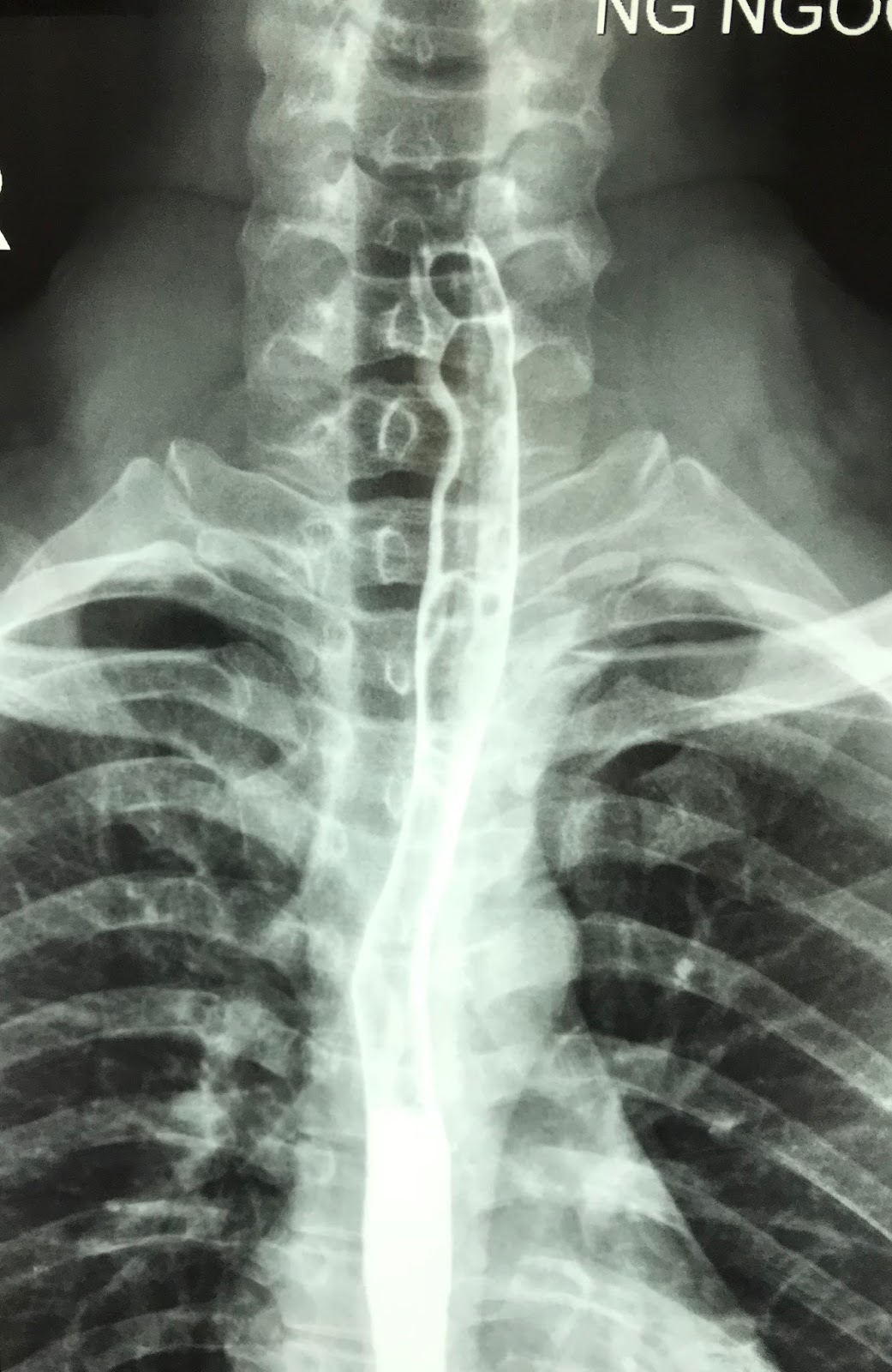
In past history he had been in an urgent operation of rupture of spleen by trauma for 10 years ( photo).


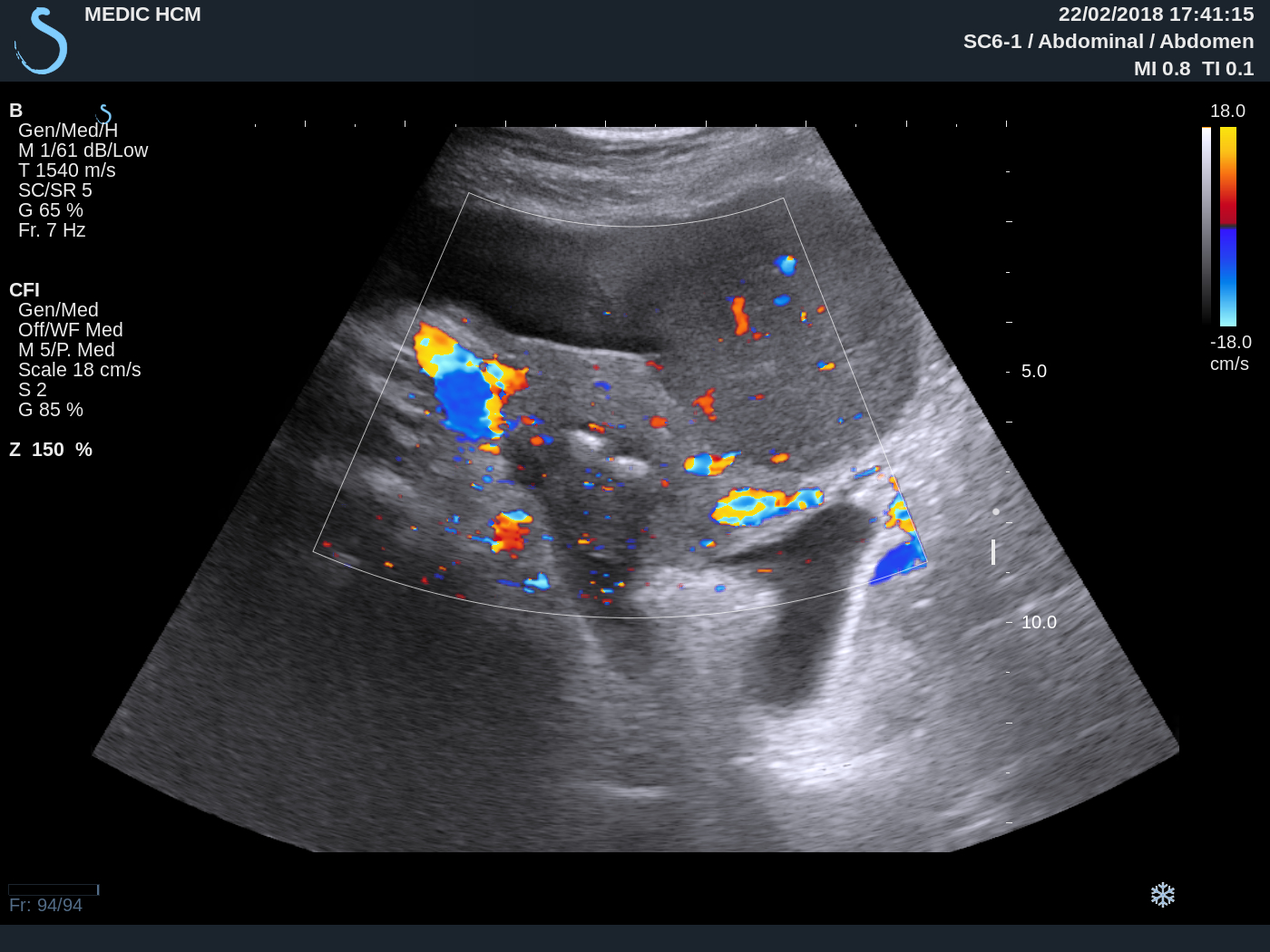
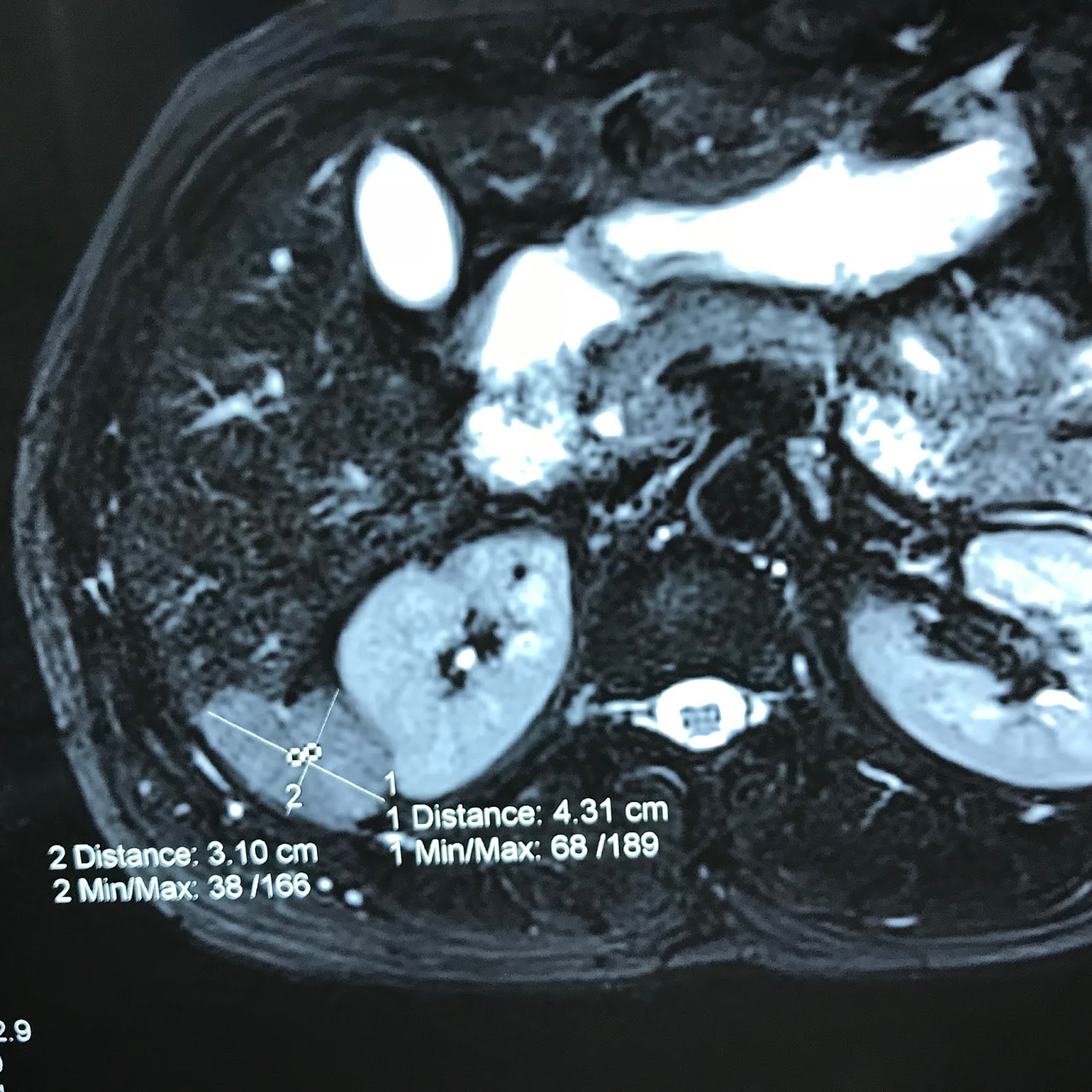
Ultrasound  detected  one  mass at border of right liver near upper pole of right kidney  and  
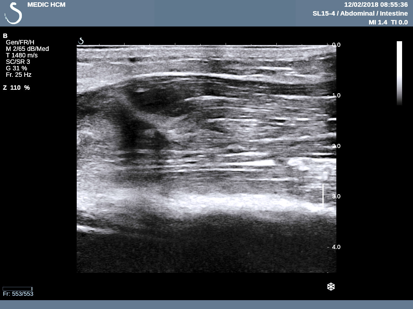
sonologist suspected an adrenal gland tumor  ( US 1, US 2 CDI , US 3  view with linear probe).


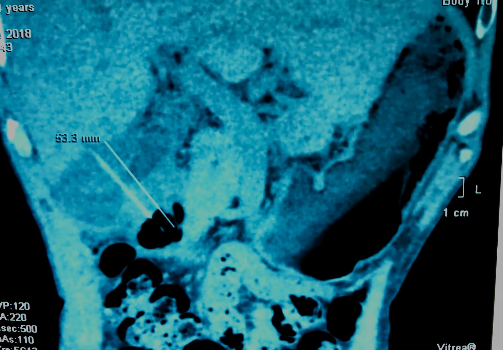
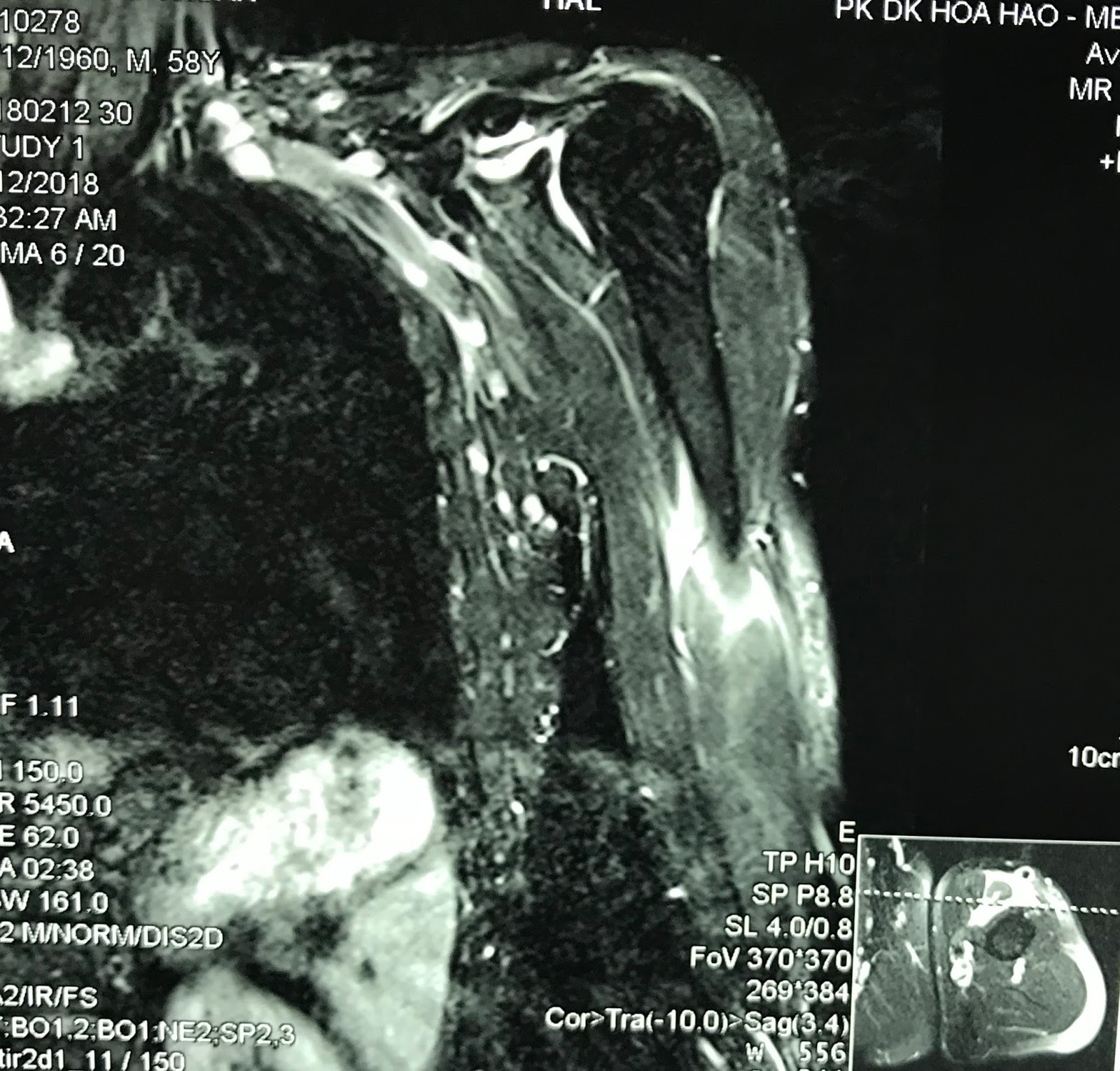
MSCT of  abdomen=   CT 1:crossed section of  tumor  at border of liver, 
CT 2 with CE  is  low enhanced  tissue.


After being treated in stable blood pressure, a laparotomy removed  big tumor at liver border in retroperitoneum  and  some intra abdomen small nodules.


Microscopic report is normal tissue of spleen.


It is  splenosis  due to rupture of the spleen 10 years before.   
DISCUSSION:
For this case  ULTRASOUND, CT, MRI  CANNOT  DIAGNOSE  SPLENOSIS.
With BLOOD TEST of WAKO NEGATIF  and HISTORY  of SPLENECTOMY, SUGGESTION pre-op IS SPLENOSIS.( MRI  WITH GADO  ALSO CANNOT  DIAGNOSE THIS CASE).


REFERENCE:

1-Thoracic splenosis  dr Nguyen quy Khoang


  2-PDF    REPORT ONE CASE .


Saturday 10 March 2018

CASE 481: ZENKER’S DIVERTICULUM, Dr PHAN THANH HẢI, Dr PHẠM THỊ THANH XUÂN, MEDIC MEDICAL CENTER, HCMC, VIETNAM.


Man  38yo with shore throat, suspected GERD but  esophago-gastric endoscopy is gastritis.


Ultrasound of the neck at left lobe thyroid detected a cyst # 1.5 cm in diameter  (US 1, US 2 ) and compression maneuver over this cyst made it smaller.


US 2:  longitudinal scanning of left lobe this cyst had air inside.
US 3:  CDI with Doppler artifact reverberation (US 4).


Sonologist report is esophageal diverticulum.
X-Rays of swallow barium showed normal esophagus.


MSCT at cervical area with iodine contrast swallow showed air in this cyst  and contrast filling inside.


Radiologist report is Zenker‘s diverticulum of esophagus.
http://www.journalmc.org/index.php/JMC/article/view/784/392

REFERENCE: Case report  of Journal of Medical Cases.


Sunday 4 March 2018

CASE 480: BILATERAL MAMMARY-OVARIAN LYMPHOMA, Dr JASMINE XUÂN, Dr TRẦN NGÂN CHÂU, Dr PHAN THANH HẢI, MEDIC MEDICAL CENTER, HCMC, VIETNAM


Woman 33yo with pain in lower part of left maxillary bone for one month and tension in both  2 breats ( see photo), hyperpigmented edema of areolar area both 2 sides without pregnancy.  


US scanning of abdomen= big cervix  of uterus,  ascites and 2 ovarian solid tumors,  size 5-7 cm (US 1, US 2). 


US of mammary scanning  showed  small  hypoechoic  nodules  infiltrating in 2 breasts without axillary node ( US 3-US 4).


US 5-US 6-US 7 =  ABVS scanning  detected  multiple nodules infiltrating in 2 breasts.


  • MRI  full body with gado detected  bone marrow changing, 2 breats  hypercaptured contrast  ( MRI 1),  ascites and kidney infiltration ( MRI 2).


MRI 3 : pelvis  with 2 ovarian tumors and big uterine cervix (MRI  4).


Blood tests=   lower platelets,  EGFR  lower  46, beta2 microglobuline raised  3816,  ferritine raised  911, LDH-l  raised  1360.
Based on clinical, imaging and blood tests  suspected diffuse type lymphoma.

Biopsy of 2 breasts  reported  microscopic with IHC, beta cell lymphoma.


Summary =  Lymphoma stage 4  infiltration in 2 breasts and 2 ovaries  for this case.  
Patient died for 4 months and 20 days from the onset of her maxillary pain.

Reference:


Wednesday 28 February 2018

CASE 479: TOOTHPICK PERFORATED BILIARY DUODENUM, Dr PHAN THANH HẢI-Dr LÊ THỊ THANH THẢO, MEDIC MEDICAL CENTER, HCMC, VIETNAM.


Man 74 yo with epigastric pain, being treated like gastric ulcer.
Ultrasound of abdomen  detected  one hyperechoic foreign body # 3.5 cm which penetrated  gastro-duodenum wall  to gallbladder. The  gallblader wall is very thick # 1 cm.


MSCT of  abdomen made diagnosis  that was a toothpick  penetrated from gastric wall to  gallbladder.


Operation removed this toothpick and cholecystectomy.


Conclusion:  Toothpick perforated gastric wall to gallbladder, an emergency case must be known.

REFERENCE: CASE 232.


Friday 16 February 2018

CASE 478: POPEYE’ SIGN of BICEPS MUSCLE, Dr PHAN THANH HẢI, MEDIC MEDICAL CENTER, HCMC, VIETNAM.


Man 58 yo playing tennis got pain in left upper arm.  Clinical  detected left biceps muscle protrusion  in contraction and painful. ( see photo 1 relaxed position,   photo 2  flexion position).


Ultrasound of biceps muscle  =

US 1 :  longitudinal scanning represented   rupture at the upper head of biceps muscle.


US 2 :  crossed section disclosed  echo poor pattern due to hematoma.  


US 3 : crossed section at middle part of biceps muscle  showed  hyperechoic pattern  by contraction.


MRI  made sure that rupture of upper part of biceps muscle.


Operation for repairing the ruptured muscle.   

Conclusion:  Orthopedic pathology due to sports named  Popeye' sign. 

Reference:   Anatomy of biceps tendon  and  Popeye’ sign.