Total Pageviews

Sunday 11 June 2017

CASE 437: LIVER TUMOR, Dr PHAN THANH HẢI, Dr VƯƠNG THỪA ĐỨC, MEDIC MEDICAL CENTER, HCMC, VIETNAM


Female patient 16 yo with epigatric pain.

Blood test=  HP positive.
Ultrasound  detected  left liver tumor.
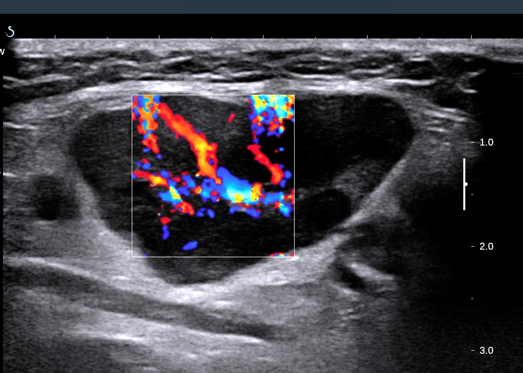
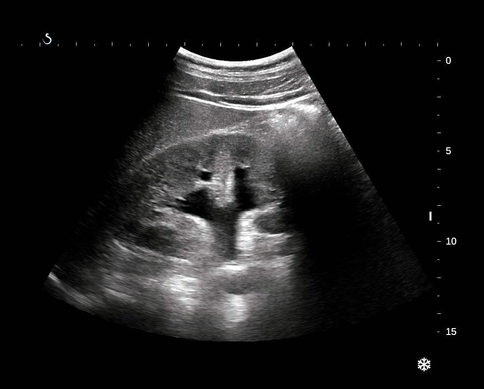
US 1 =  longitudinal scanning of  left lobe of liver:   solid tumor , size  of 10 cm with central necrosis.

US 2 =  subcostal scanning :   tumor covers left liver lobe.

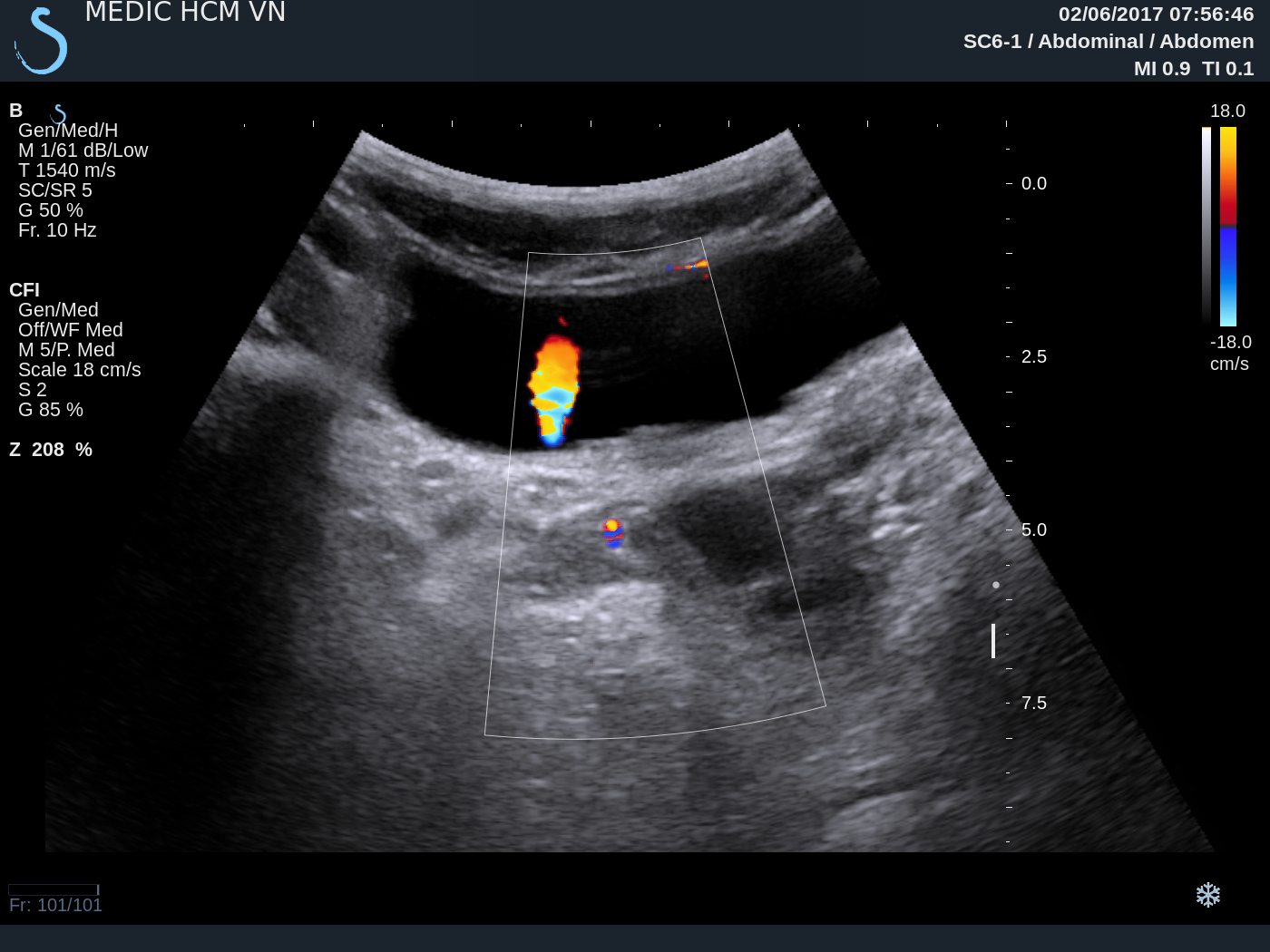
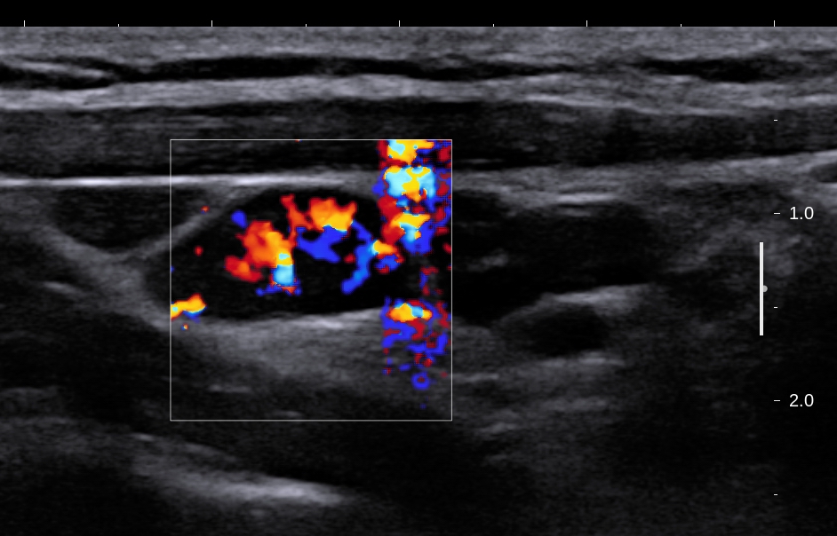
US 3  = color Doppler :  vascular supplying  of  this tumor.


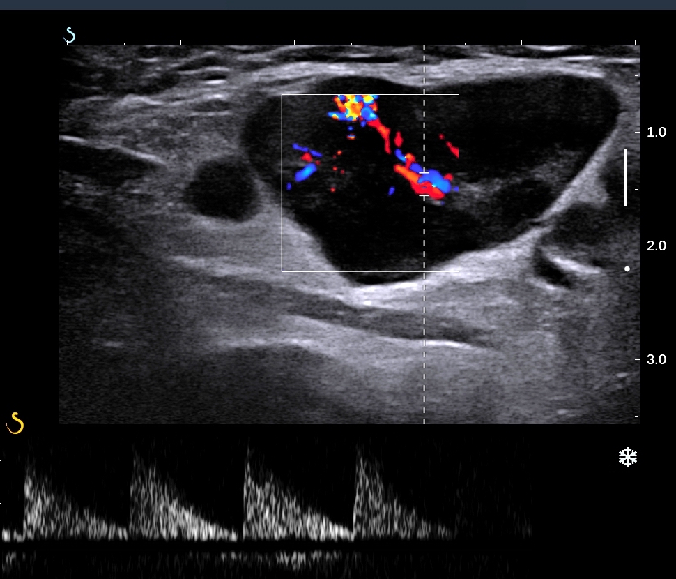
US 4 =  elastoscanning of this tumor = 10,5-15 kPa.  


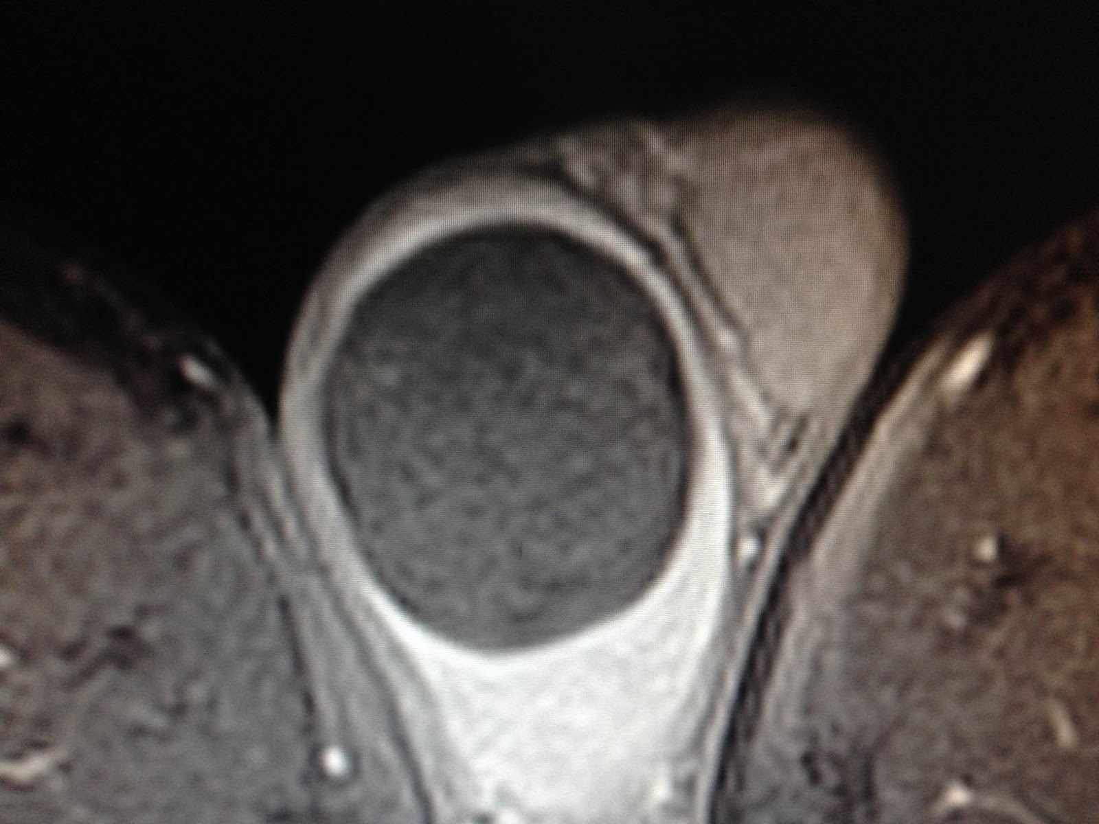
MSCT with  CE: CT 1, CT 2 =  artery and vein phases, CT 3 =  frontal view of this tumor from left liver.


Blood tests=  no infested HBV, HCV, Wako tests 3 negative.


PRE OPERATIVE  RADIOLOGIST AND SURGEON  SAY THAT  LIVER FOCAL NODULAR HYPERPLASIA (FNH).

OPERATION WAS DONE FOR RESECTION OF THE TUMOR (SEE  MACRO).


Macroscopic is  FNH.


Liver tumor FNH or HCC


Reference: Macro FNH and  flow chart of identification FNH-HCA-HCC.


Friday 9 June 2017

CASE 436: SOLITARY KIDNEY, Dr PHAN THANH HẢI, MEDIC MEDICAL CENTER, HCMC, VIETNAM


Man 32yo  with acute pain at  right renal fossa.
Emergency ultrasound:
US 1 = right kidney  hydronephrosis, no left kidney.
US 2 =  crossed section of  right kidney.
US 3 =  right  ureter dilated and stone of 1.5 cm in diameter.
US 4 = color Doppler  twinkling artifact  with stone.


US 5 = in  urinary bladder , urine jet only on right site.


US 6 =  cystic mass at the left wall of urinary bladder.


MSCT with CE=  
CT1:  fron tal view,  right  kidney  hydronephrosis no left kidney.
CT 2:  sagital view   the stone in right ureter.

CT 3:  3D view.
CT 4:  vascular malformation of agenesis of left kidney.


Saturday 3 June 2017

CASE 435: CERVICAL LYMPH NODES, Dr PHAN THANH HẢI, MEDIC MEDICAL CENTER, HCMC, VIETNAM


Female patient 23 yo detected  submandibilar nodes with history of recurrent sore throat. And some palpable  left cervical  nodes along  of SCM. No fever, nor pain.
US 1: hypoechoic, no hilus left submandibular nodes.


US 2: scanning  along  left site SCM, small hypoechoic nodes.


US 3: hypervascular  color Doppler .


US 4. high flow vascular  intranodes without defected area.


US 5: spectral Doppler  high RI and PI.


US 6: elastoscanning of  inhomogeneous nodes, central low kPa.


Biopsy of  the big submandibular node.
Blood tests=  normal WBC, CRP,  but high beta microglobuline 3200 UI, and  normal LDH, ferritine.

Microscopic report of this biopsy is tuberculosis  lymph nodes with LANGHANS  MACROPHAGE CELLS.


SUMMARY :TUBERCULOSIS CERVICAL LYMPH NODES.

Monday 29 May 2017

CASE 434: TESTIS TUMOR, Dr PHAN THANH HẢI - Dr LÊ VĂN TÀI, MEDIC MEDICAL CENTER, HCMC, VIETNAM.


Man  36yo with right testis tense.
Ultrasound scanning of  right scrotum  detected big testis focal lesion,  round,  size of 3cm
US 1  color doppler  not  hypervascular, well bordered.

US 2 CDI: hypovascular tumor.

US 3  elastoscan of  this tumor is inhomogeneous with some parts very hard.


MRI  with gado: MRI 1, 2, 3: this tumor  very low gado enhanced.


Radiologist suggested epidermoid  cyst.
Blood tests =  normal   AFP  and   HCG.
Operation   resection of  right testis (see  macro 1, 2).


Microscopic report is epidermoid cyst.


Tuesday 23 May 2017

CASE 433: GIANT RETROPERITONEUM LIPOMA, Dr PHAN THANH HẢI, MEDIC MEDICAL CENTER, HCMC, VIETNAM.


Woman 44yo nullipareous, with abdomen  distention, and  clinical  suspected   ovary tumor.
US scanning of abdomen detected  all bowell loops in deplacement to left flank.

US 1: epigastric scanning detected  right kidney near left lobe liver.


US 2: big solid mass, hyperechoic  like  fatty tissue.


US 3, US 4  color Doppler  of this mass showed from  retroperitoneum which extended from the epigastric  to pelvis but  no deplacement of abdominal aorta.


MRI  with  gado:  This  big mass is in retroperitoneum deplaces right kidney to liver (MRI 1, MRI 2, MRI 3,  MRI 4 , MRI 5).  


Radiologist reports  that  retroperitoneal lipoma or retroperitoneal liposarcoma.
Operation removed this mass easily (see macro).


Microscopic report is lipoma.
Reference:

Sunday 21 May 2017

CASE 432: A K I by STONE, Dr PHAN THANH HẢI, MEDIC MEDICAL CENTER, HCMC, VIETNAM


Man  51 yo with acute  pain at  right flank, type colicky  pain. History of being treated  renal stone of  left kidney by operation and ESWL for 2 years.
Emergency  ultrasound  detected right and left kidney hydronephrosis (US 1, US 2).


CT scanning  with CE:  CT 1= kidneys no CE.


CT 2 with CE,  arterial phase.


CT 3,  venous phase.


CT 4,  delay phase.


CT 5,  frontal view.


 CT 7,  3D view.


Blood test: EGFR=  23mL/s.

Discussion: Ultrasound  scanning in acute renal colic crisis cannot make diagnosis of  A K I ( acute kidney insufficiency);   CT  non CE  with HU  low and CE phase in delay  secretion that suspected AKI.

 Emergent  operation was done  for removing  of the stuck stone in right ureter for this case.