Total Pageviews

Wednesday 10 May 2017

CASE 429: DOUBLE UTERUS, Dr PHAN THANH HẢI- Dr TRẦN NGÂN CHÂU, MEDIC MEDICAL CENTER, HCMC, VIETNAM.

Woman 48yo, PARA 2002 , detected herself one prolapsed mass from her vagina 1 year ago, no pain, no fever but with SUI [ stress urine incontinence] syndrome.


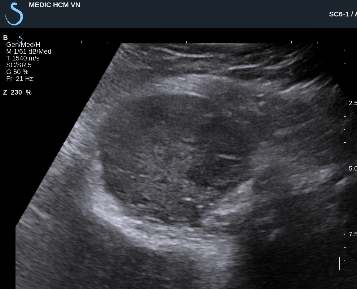
Ultrasound of pelvis by the transcutaneous: (US1) uterus normal size,


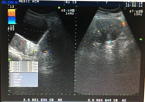
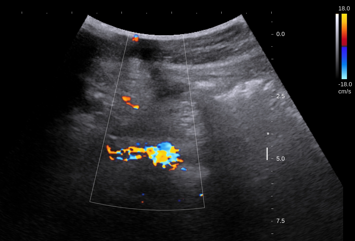
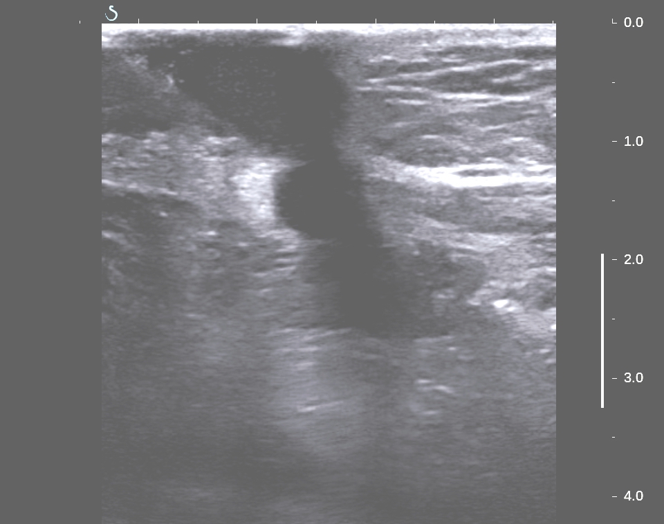
by via TVS ( US2, US 3) detected one mass at lateral left uterus, hypoechoic, size 7 cm look-liked second uterus.


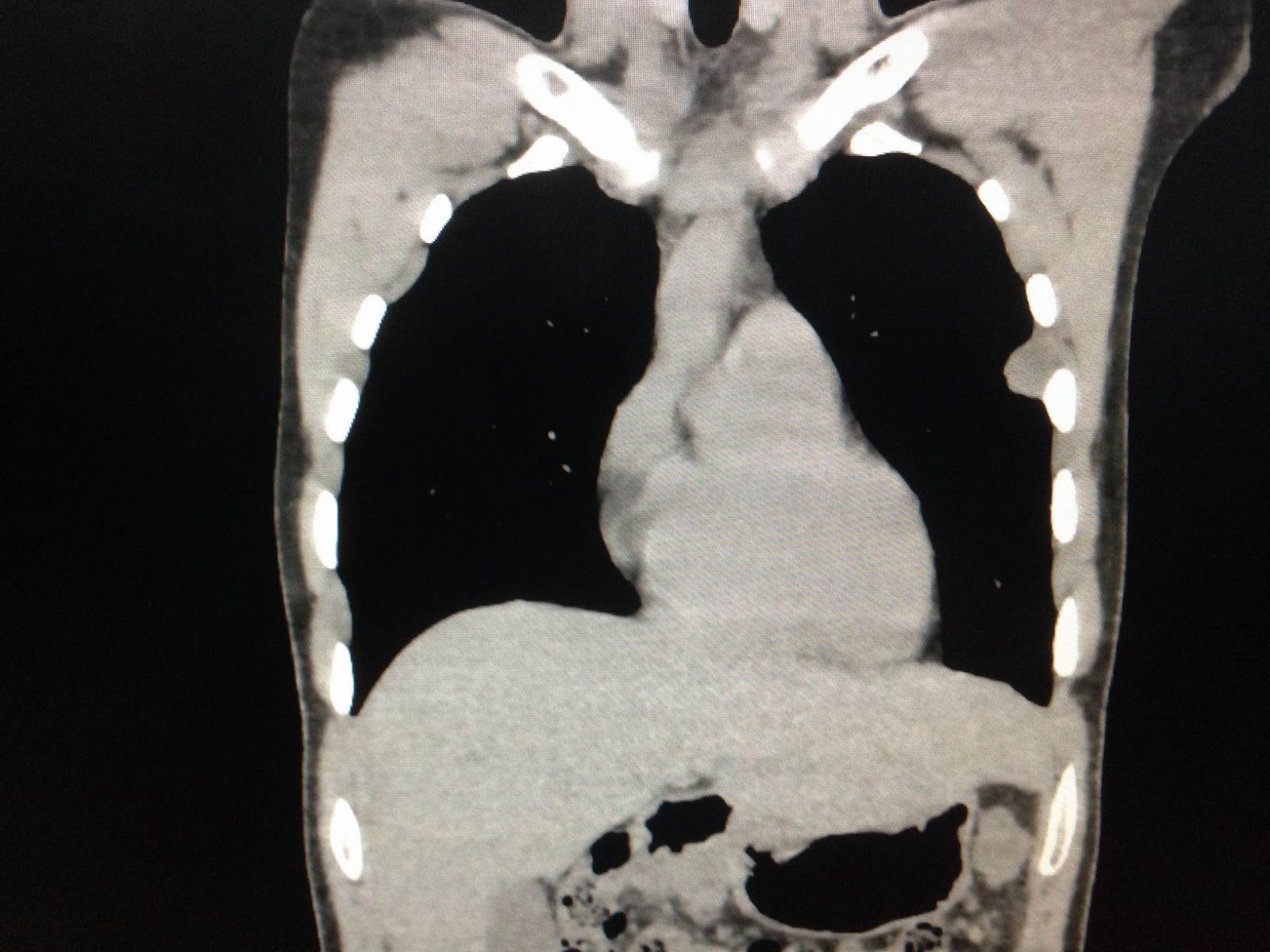
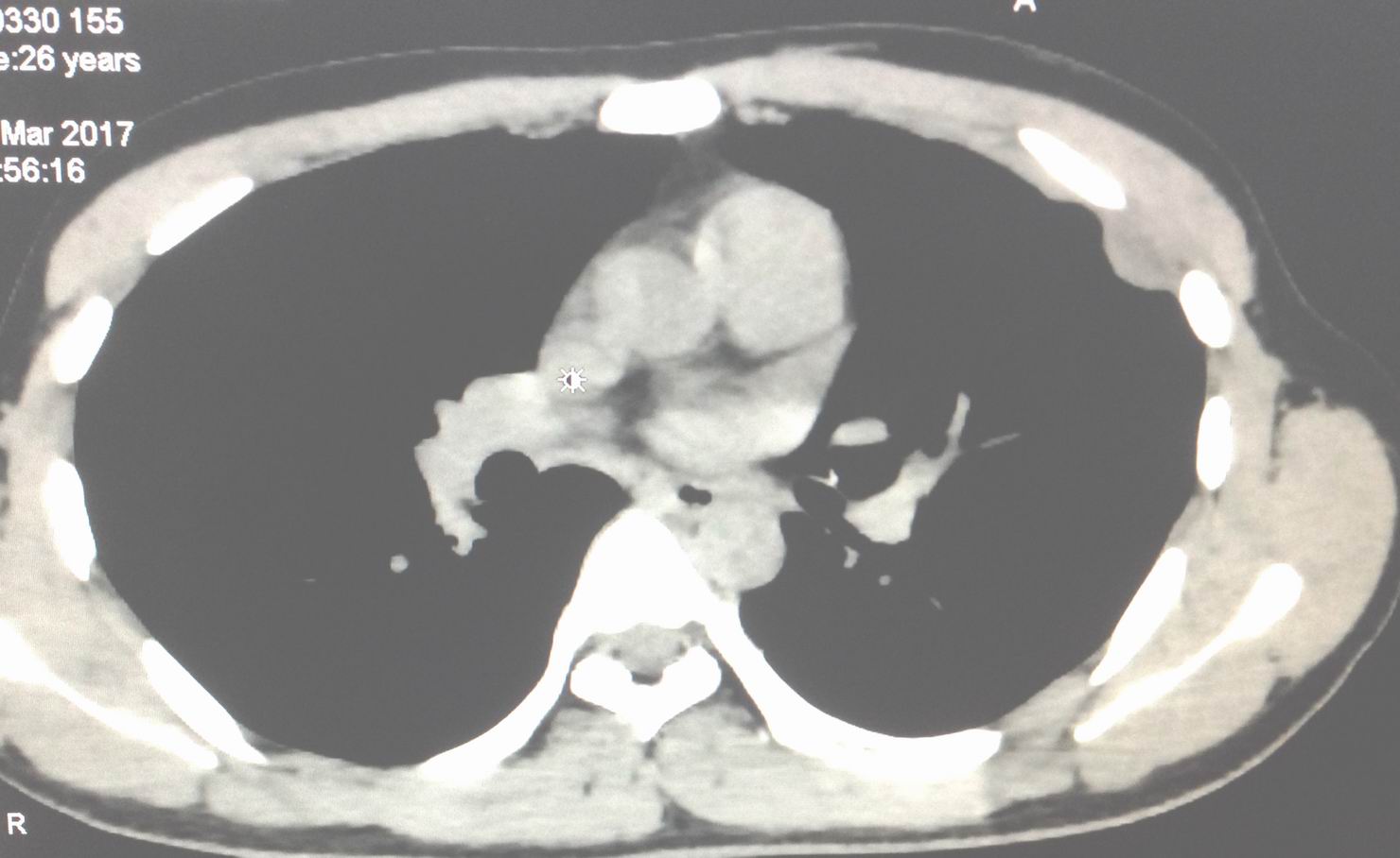
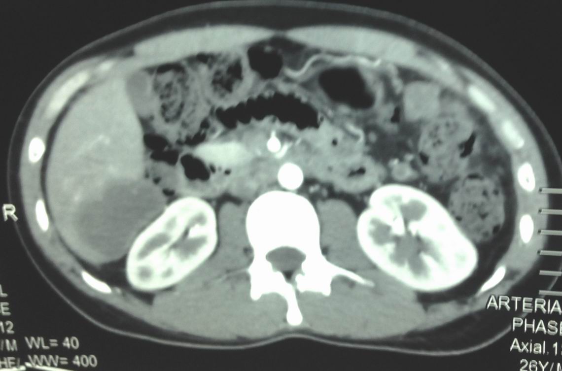
CT scan of pelvis:  CT 1 : this mass at left site uterus, hypodense like fatty tissue.


CT2, CT3 the mass is anterior the urinary bladder.


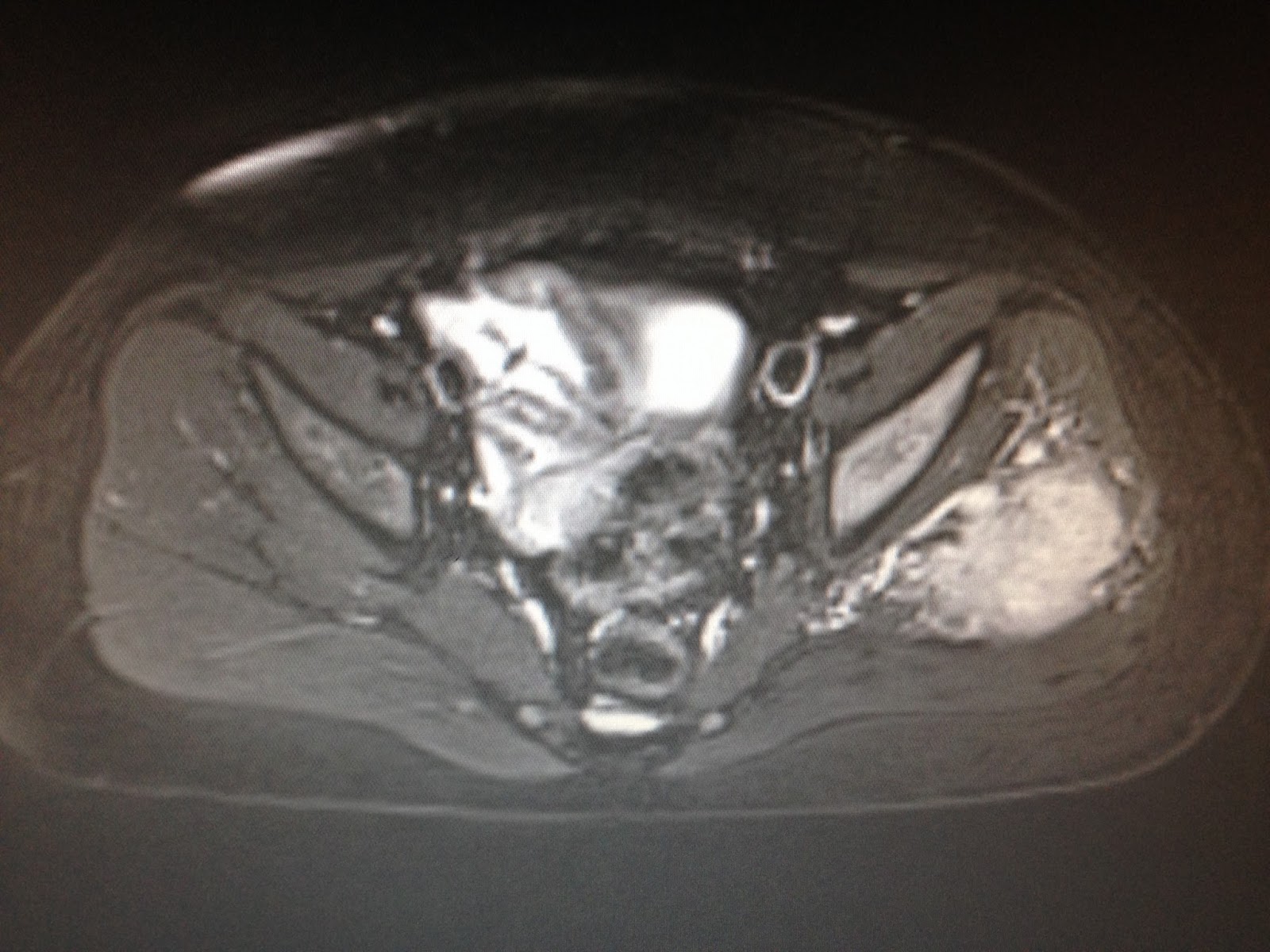
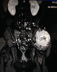
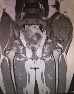
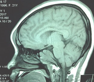
 MRI 1, MRI 2 in sagittal section, this tumor is like a second uterus.


OPERATION  REMOVE  THIS TUMOR..BY LAPAROTOMY..
( OP. IMAGES:THIS TUMOR  IS  NOT  FROM UTERUS)


MICROSCOPY IS FIBRO-LIPOMA  

Saturday 15 April 2017

CASE 428: BUTTOCK TUMOR ASPS, Dr PHAN THANH HẢI, MEDIC MEDICAL CENTER, HCMC, VIETNAM


8yo female child detected one mass at left buttock that was in slow growth for 3 month and getting  pain now.
Ultrasound of this mass which is  located at gluteus medius muscle of left buttock.


US 1: Hypoechoic mass, size of 5 cm, well bordered.


US 2 : CDI, hypervascular color mapping looked like a ring.


US 3: PWD, RI low of  the artery supply.

MRI  with gado of the mass.


MRI 1, MRI 2, MRI 3 of  this mass  showed  erosion of the iliac bone  and  MRI 4 revealed a small mass intraspinal canal.


Open biopsy of  this tumor with  the specimen  looked like  brain tissue and microscopic result is ASPS  (alveolar soft part sarcoma).


REFERENCE: CASE REPORT.


Monday 10 April 2017

CASE 427 : POLYCYSTIC BREAST, Dr PHAN THANH HẢI, Dr JASMINE THANH XUÂN, MEDIC MEDICAL CENTER, HCMC, VIETNAM


Woman 27 yo  with history of the left polycystic  breast detecting by herself since April 2016 .
 FNAC reported  nothing  abnormal detected, and she went to Medic for 3 times [each in 3 months]
 with the same result of polycystic left breast without tumor.


 But now she got pain at left breast and decided to reexamination.

Ultrasound of the left breast in the 4th examnination showed many small simple cysts but one   of them  is biggest
 with  size of 3x 4 cm.  At later time, the biggest cyst with thin wall but  having one   hypervascular 
vegetation mass, size #1.5 cm.


 US 1:Big cyst with  fine septation.


US 2 :  Small simple cyst.


US 3: Intracystic mass.


 US  4: CDI  hypervascular mass.


Strain elastography of intracystic mass showed a stiff area (mixed pattern)
which was corresponded with a score of 2 (Tsukuba score).


FNAC again with no abnormal cell,  only red blood cells.


Liquide analysis: no  abnormal  of markers  CEA, CA 125, CA 15-3.

  ABVS ( AUTOMATIC  BREAST  VOLUME  SCANNING) shows the  intracystic tumor
  by 3D VIEW.


  Operation for removing this big cyst (see macro).


Microscopic report is  benign cyst with  intracystic papilloma.


Thursday 6 April 2017

CASE 426: MULTIPLE TUBERCULOSIS ABSCESSES, Dr PHAN THANH HẢI, MEDIC MEDICAL CENTER, HCMC, VIETNAM


https://mail.google.com/mail/u/0/images/cleardot.gif
Male 26yo with umbilicus swelling and pain.
Abdominal ultrasound   detected  abscess of umbilicus  ( US 1, US 2), liver abcess and  left pleural  abscess.


MSCT  confirmed  abscess  of  left pleural, liver and umbilicus.


Blood tests:   WBC 12k, CRP  normal.


Punction of the umbilicus abscess  withdrawed  white thick pus, high ADA test :104 UI/mL
Conclusion: it is multiple abscesses due to tuberculosis.

Reference: ADA.pdf


Thursday 30 March 2017

CASE 425: FRONTAL BONE TUMOR, Dr PHAN THANH HẢI, MEDIC MEDICAL CENTER, HCMC, VIETNAM


Woman 21 yo with headache, blurred vision and protrusion of frontal area of face  (see photo) for 3 months.


Ultrasound of  frontal area  and eyes detected  hypoechoic  and hypervascular mass which made destruction the frontal bone, but the  orbit  remains intact, while periorbital part was infiltrated by a  hypoechoic structure ( US 1, US 2, US 3).


MRI of the brain and cervical column revealed no intracerebral tumor and the cervical  bone changing structure but not destruction. There is erosion of frontal bone  with mass  under skin of the frontal area.  


Blood test showed very high beta 2 microglobulin.


Suggestion for this case is multiple lymphoma with infiltrating of frontal area.

Reference of a case on  large B-cell lymphoma of the Frontal Bone.