Total Pageviews

Monday 26 September 2016

CASE 396: PARATHYROID CYST, Dr PHAN THANH HẢI, Dr NẠI HƯƠNG THOANG, Dr VŨ TU THÂN


Woman 43 yo with  sorethroat  and cough, sputum bleeding.
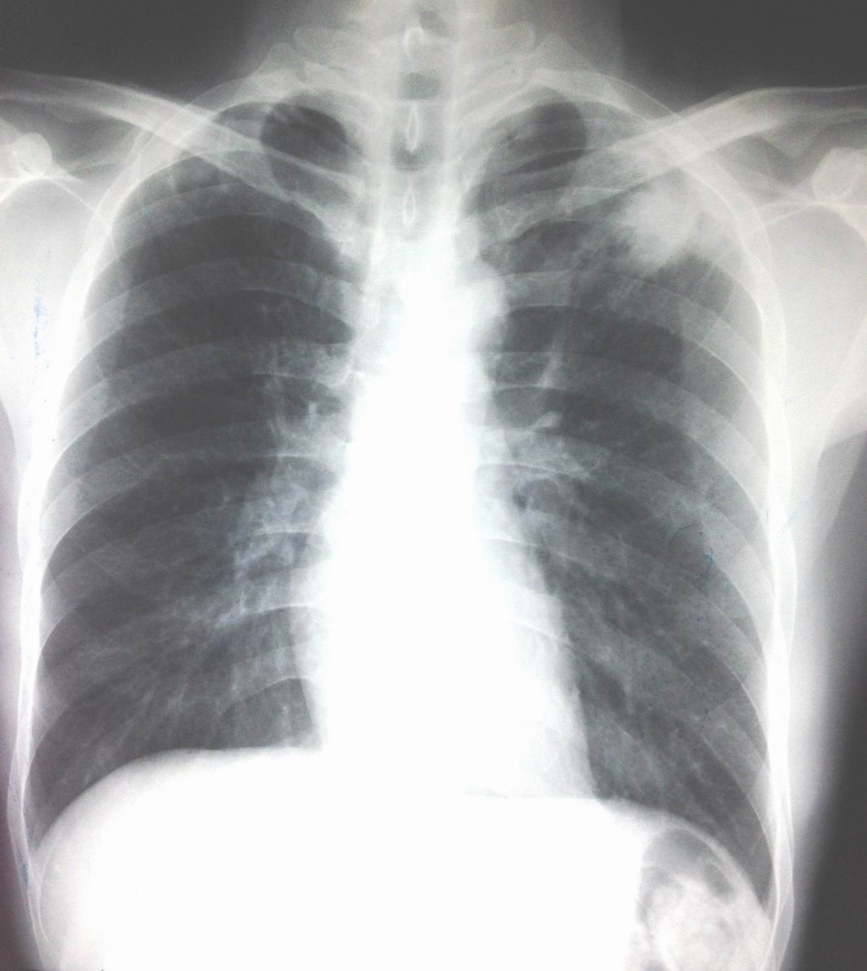
Chest X-Rays for  screening: no  chest lesion ( see foto chest X-Rays).


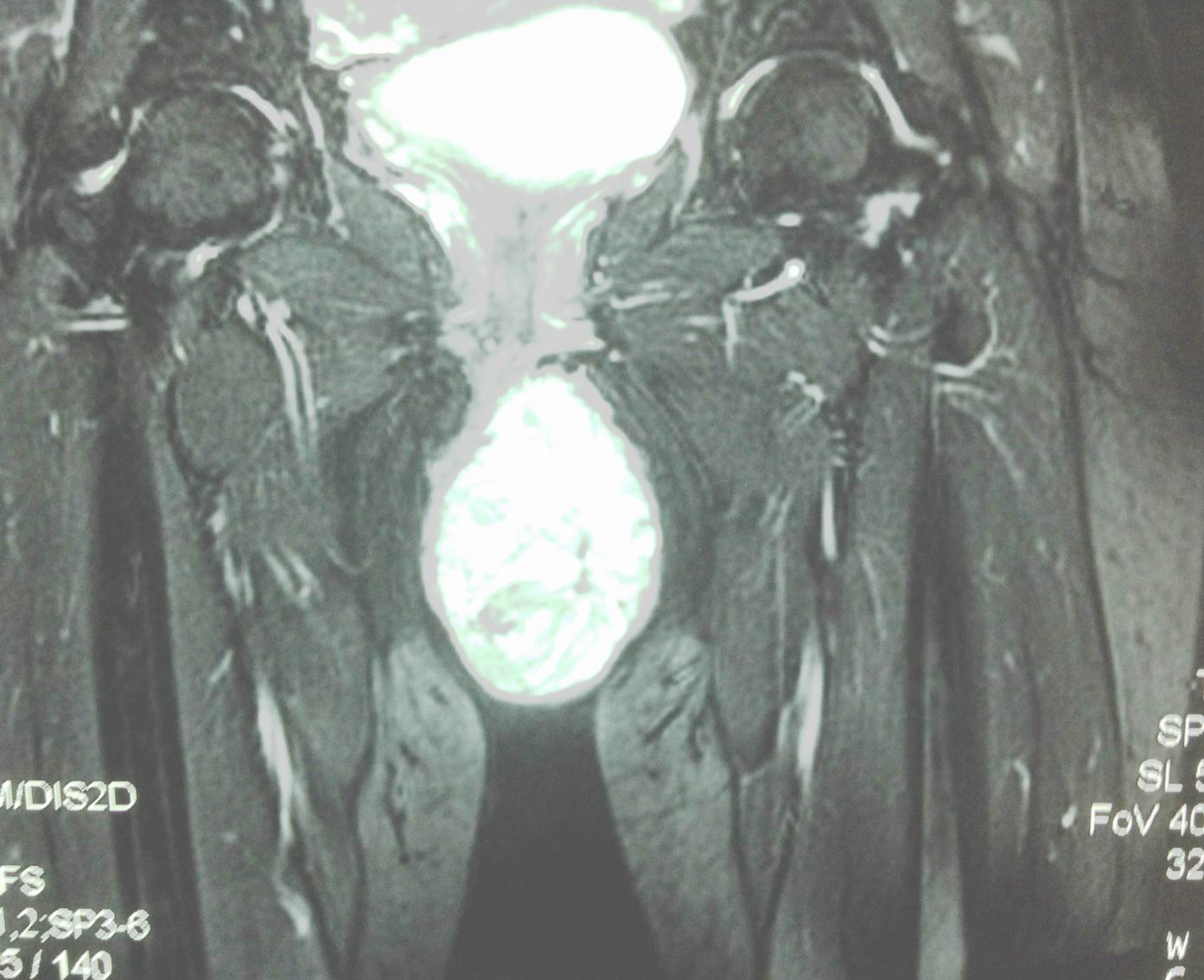
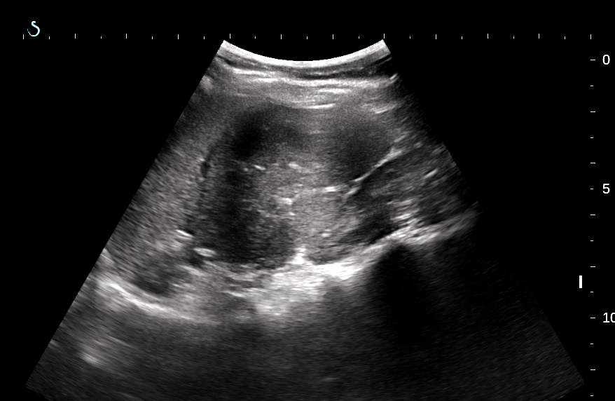
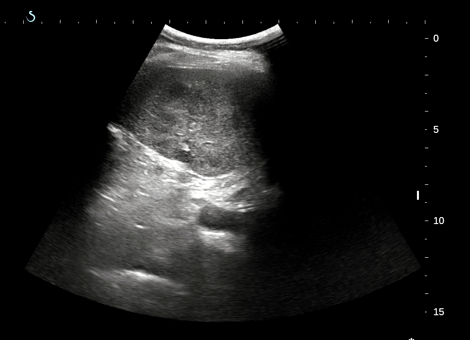
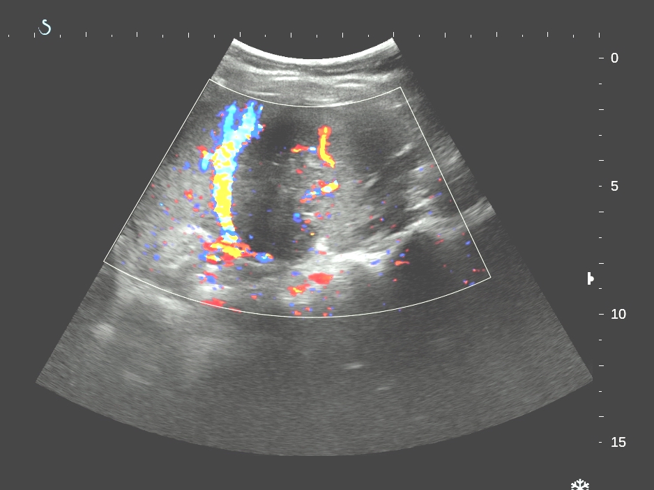
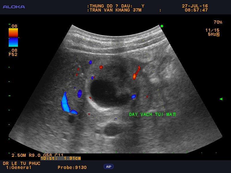
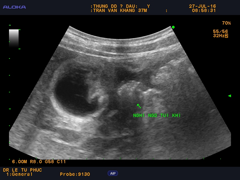
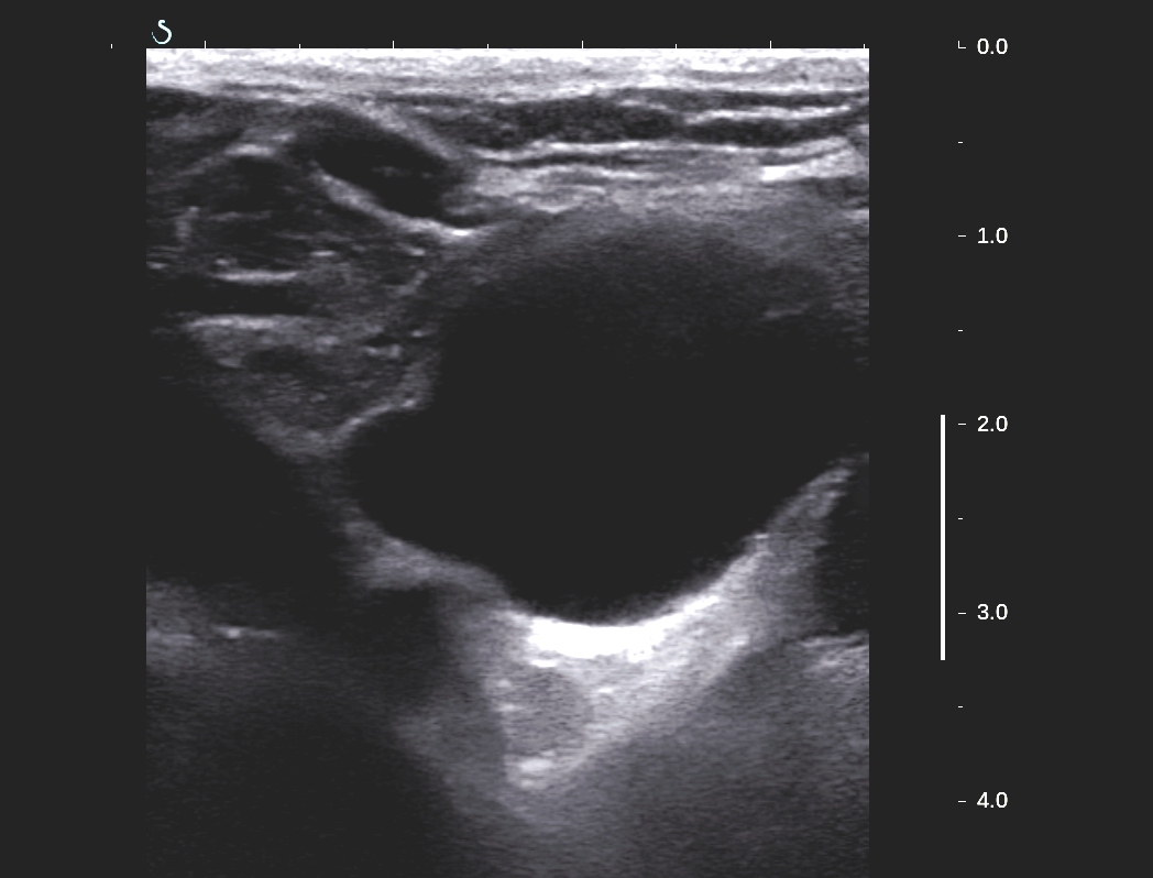
Ultrasound of  the neck: normal thyroid  but detected a cyst at lower pole of thyroid gland, size of 5-6 cm, monocystic  prolonged to retrosternum.
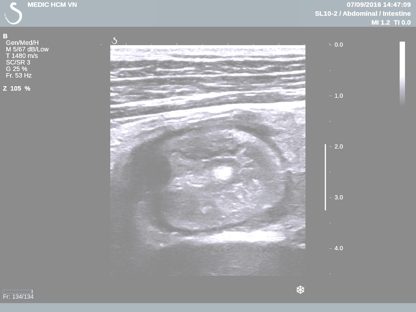
US 1,US 2 ( CDI),   US 3    pretrachea longitudinal scanning.


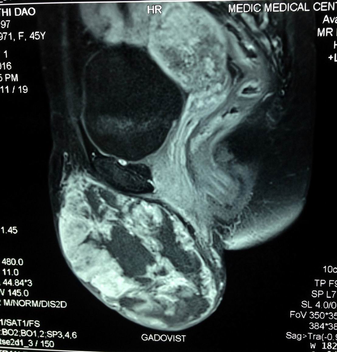
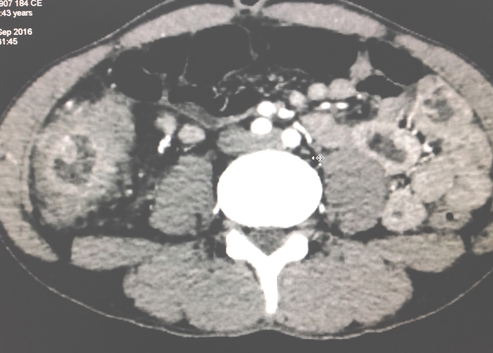
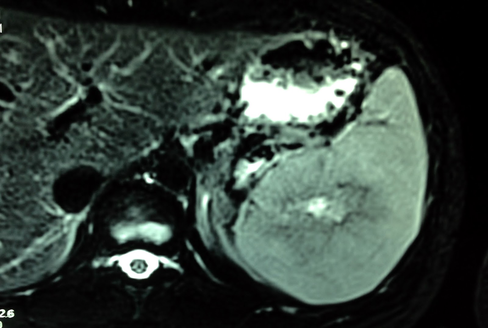
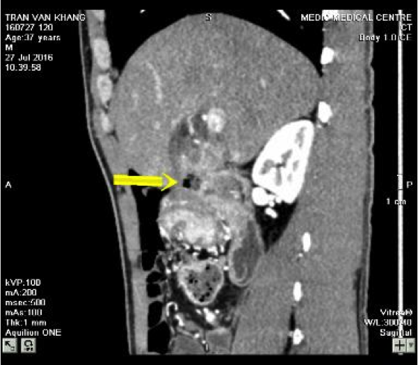
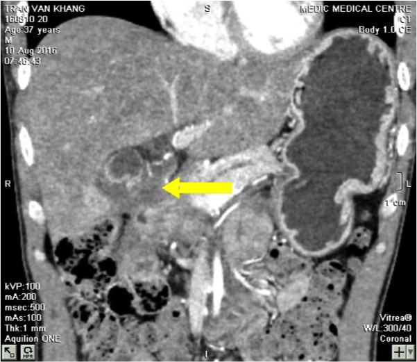
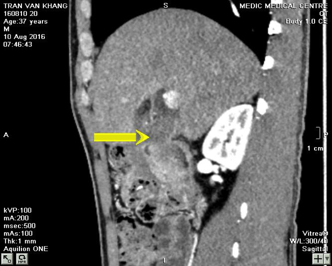
MSCT CE of  the neck: CT 1=frontal viewing,  well bordered cyst, CT 2: sagittal view..
C T 3. Cross-section= retrosternum tumor.


Ultrasound guide punction  of this cyst removing 10ml clear fluid.
What do you need to study in this fluid?

Fluid analysis report: high PTH  78.69pg/ml  and in the blood PTH is 47pg/mL.

The final diagnosis is  non functional PARATHYROID  CYST.

  
Reference


Monday 19 September 2016

CASE 395: MAJOR LABIA TUMOR, Dr PHAN THANH HẢI, MEDIC MEDICAL CENTER, HCMC, VIETNAM


WOMAN 45 YO, SAYS  THIS VULVA MASS  IN  SLOW GROWING 6 MONTHS AGO, WITHOUT  PAIN ( FOTO).


ULTRASOUND OF THIS TUMOR REVEALED SOLID, FIRM,   SIZE OF 10CM.
 US 1:WITH  B MODE,    US 2 WITH  CDI : HYPOVASCULAR SOLID TUMOR /  US 3=ELASTOULTRASOUND OF THIS TUMOR IS HARD STRUCTURE.


MRI 1 SCAN = SAGITTAL SECTION OF THIS TUMOR
MRI 2 WITH  GADO  CE= IN LATE PHASE, LOOK LIKE  A CYSTIC DEGENERATION.
 MRI 3 = FRONTAL  SECTION OF THIS MASS  FROM LEFT MAJOR LABIA,  PEDUNCULATED  LOOK  LIKE SCROTUM IN MALE PATIENT.


Core  biopsy reported  microscopic with  immmunochemistry staining is  glomus tumor.


Operation remove this tumor (see macro)


.
Reference:

https://www.scitechnol.com/glomus-tumor-in-vulva-with-uncertain-malignant-potential-1Mbz.pdf

Tuesday 13 September 2016

CASE 394: COLON TUBERCULOSIS, Dr PHAN THANH HẢI, MEDIC MEDICAL CENTER, HCMC, VIETNAM


Man 43 yo with epigastric pain  crisis and  gastroendoscopy showed  gastritis.
Ultrasound  detected  one mass  like target  with  thickening of the  wall of colon (see  US 1=csoss-section  colon over  right kidney);   US  2  with linear  probe= colon wall is thickening; US 3, US 4 =  longitudinal scan).


MSCT of abdomen with CE revealed  thickening of  ascending colon wall  (CT1,  CT2).


Chest X-rays  before endoscopy  detected  infiltration of  left upper lung.


Colonoscopy reported the mass in right colon, nodular ( see foto) biopsy. 


Report of endoscopist is colon cancer.
Microscopic report  is colon tuberculosis.
Conclusion = this  case  represented colicky pain at epigastric region but  ultrasound and CT  suggested  colon cancer, same as colonoscopy, but  microscopic is tuberculosis.of colon and left lung.

REFERENCE:


Sunday 4 September 2016

CASE 393: SPLEEN TUMOR, Dr PHAN THANH HẢI, MEDIC MEDICAL CENTER, HCMC, VIETNAM


Woman 26 yo with no clinical symptom. Ultrasound screening detected a spleen mass
US 1= longitudinal scan of this mass size of 6.0cm at lower pole of spleen, hypoechoic, well bordered.

US 2=cross-sectional view of mass.


US 3=CDI of this mass with vascular bending sign and, ( US 4) structure inside hypervascular.


Blood tests are normal.
CT with CE:CT 1 non CE , CT2 CE, delay phase with central mass lower perfusion.


MRI with gado: this tumor is well bordered, peripheral enhanced and central hypoperfusion at the late phase.


LAPAROSCOPY  FOR  SPLENECTOMY  . SURGEON REPORTED  THIS TUMOR IS INTRA SPLEEN AND ITS COLOR  LOOKS LIKE SPLEEN TISSUE ( SEE  SPECIMEN).HE SAID IT MAY BE  HEMANGIOMA.

MICROSCOPIC REPORT  IS CAVERNEOUS HEMANGIOMA.

Monday 29 August 2016

CASE 392: PERFORATED SEAL-OFF DUODENUM, Dr LÊ TỰ PHÚC-Dr PHAN THANH HẢI. MEDIC MEDICAL CENTER, HCMC, VIETNAM.


"A 37-year-old male presented in our hospital with moderate epigastric pain for three months. Around one week before examination, the pain became more severe, but he didn’t recognize a suddenly pain. Clinical examination revealed no muscle guarding and rebound tenderness.

Abdominal ultrasound images showed unconcentric wall thickening of the gallbladder. Beside the more thickening wall of gallbladder, a hyperechoic of air collection was found. This air collection was continuous with small hyperechoic air spots inside duodenum. A perforated duodenal ulcer with air leakage was suspected.


CT-Scan confirmed air collection beside a thickening gallbladder wall.


Blood test indicated and raised of white blood cells (10,350 / mL) with low level of CRP (0.9 mg/L) and possitive Helicobacter Pylori test.

Without surgery, the patient pain released and the air collection was disappear in ultrasound and CT-Scan images for one month follow-up. Gastroduodenal endoscopy showed a healing ulcer in the anterial wall of duodenum. White blood cell count returned to normal."


This is a case of perforated seal-off duodenum revealed by ultrasound and confirmed by CT-scan later and successfully management without surgery.