Total Pageviews

Wednesday 11 December 2013

CASE 225: LEFT RENAL MASS, Dr PHAN THANH HẢI, MEDIC MEDICAL CENTER, HCMC, VIETNAM


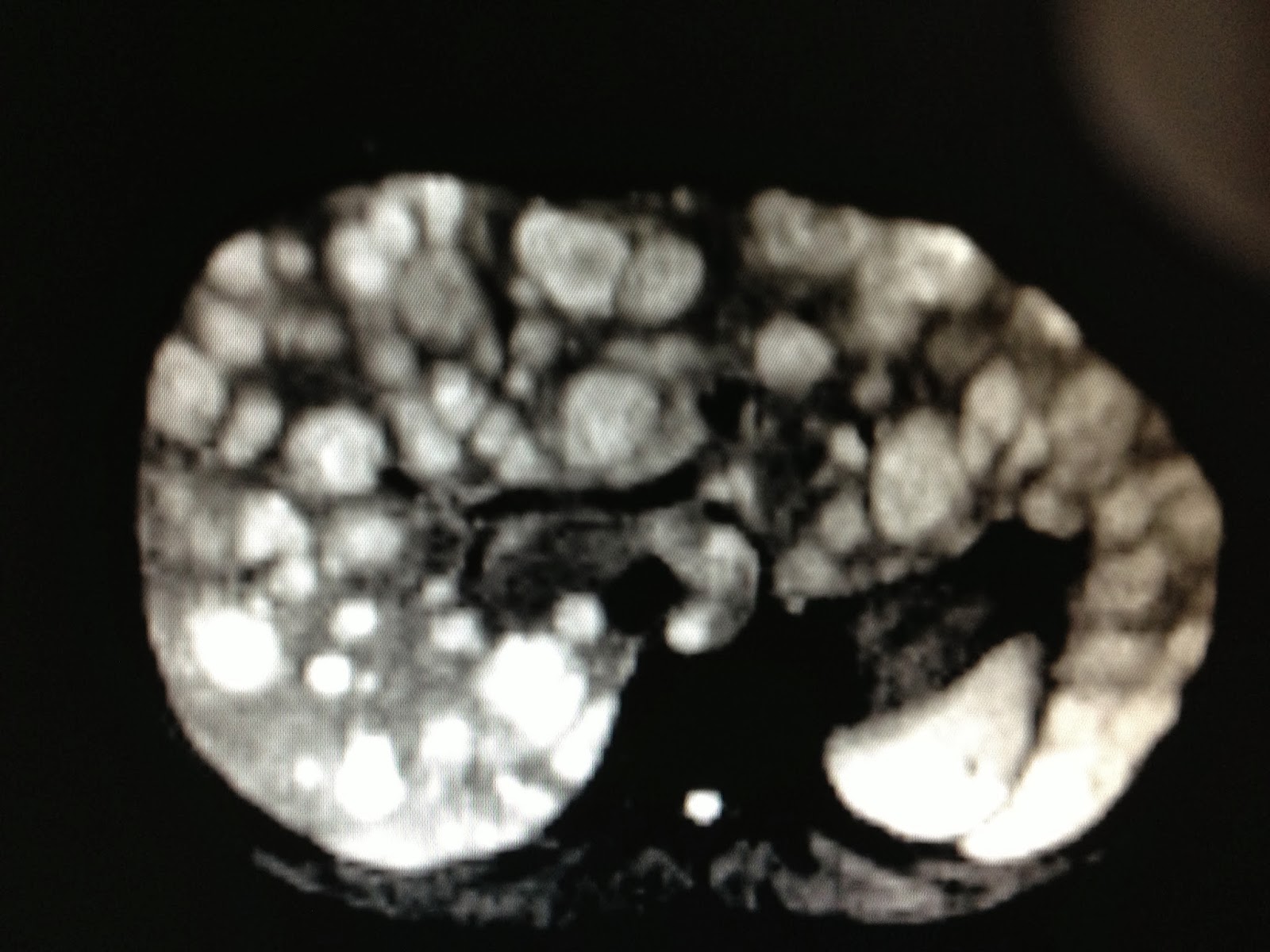
Woman 32 yo in health care check-up:  ultrasound of abdomen detected  left renal mass in hyperechoic central  kidney with size of 2.5 cm, irregular border, and  hypovascular. Elastoscan  showed that hard mass.


MSCT with CE of this mass is in rapid enhancement and quickly washed-out.


What is your suggestion?
THIS CASE MUST BE BIOPSY OR NOT?
WHY OPERATION IS DONE; LEFT LAPOROSCOPIC NEPHRECTOMY
SEE MACROSCOPIC SPECIMEN.


Discussion:
Why we do not biopsy this case, see Ref. 2009-AUA guideline


The  microscopic report  of histo-immunostaining  is AML.


Friday 6 December 2013

CASE 224: LEFT SUPRACLAVICULAR MASS, Dr PHAN THANH HẢI, MEDIC MEDICAL CENTER, HCMC, VIETNAM


Man 41 yo, cough  and  pain at left arm; chest  x-rays suspected lung tuberculosis, then he had been treated as antituberculosis  for 3 months. 


But the pain was getting worse and  one mass appeared at  left supraclavicular  fossa.
X-rays  again  showed this  first rib in erosion on the left site.


Ultrasound scanning at left supraclavicular fossa and disclosed a solid  mass in calcification


 and fluid collection on the right site.


MSCT with  CE  of the lungs detected the mass destructing the first rib. 


Biopsy was done.  

Biopsy  microscopy report  is   adenocarcinoma , it is PANCOAST TOBIAS  SYNDROME.

Ref  PANCOAT -TOBIAS SYNDROME:


History photo of  Dr  PANCOAST K Henry.

Sunday 1 December 2013

CASE 223: BIG IVC, Dr PHAN THANH HẢI- Dr NGUYỄN KIM THÁI, MEDIC MEDICAL CENTER, HCMC, VIETNAM


MAN  33 yo  PECTUS EXCAVATUM,  ONSET OF  EPIGASTRIC PAIN  WITH  RADIATION  TO BACK  DORSAL SPINE.


ULTRASOUND OF ABDOMEN  DETECTED  BIG IVC  WITH  SIZE  OF =2.4-2.7cm,  ECHOGENIC  BLOOD FLOW.


SPECTRUM DOPPLER OF IVC SHOWED TRIPHASIC PATTERN WITH PV  11.2cm/s.


TEE 3D  CARDIAC  REPORT WAS NORMAL.


WHAT IS  CAUSE  OF BIG  IVC ?

MSCT of abdomen showed that IVC  is dilated in  going to heart,  diameter of 2.9-3.3 cm.

What is your suggestion ?.

REFERENCE  IN INTERNET OF  THE CASE ABOUT PECTUS EXCAVATUM AND IVC IS SAME AS  THIS CASE,  SUGGESTED  IVC  DILATED  DUE TO  PE (PECTUS EXCAVATUM).


Thursday 28 November 2013

CASE 222: LIVER MULTIPLE SPOTS due to COLON CANCER, Dr PHAN THANH HẢI, MEDIC MEDICAL CENTER, HCMC, VIETNAM


Man 31 yo  onset fever one month ago but not control by antibiotics. Pain in liver region.
Ultrasound  detected  some round spots  like  liver abscesses.(US  B mode, CDI, Elasto).


MRI  with  gado CE detected  also  many  round  spots,   same size of  2 cm, with central necrosis looked like metastasis.


Blood tests=   WBC rising of 20k with 85% neutro, negative sero amibe, normal CEA.


Colonoscopy for detection of primary cancer disclosed, at splenic angle of colon, infiltrated induration lesion which was suspected colon tumor .


 And biopsy with microscopic report was colon cancer.


Summary; this case is colon cancer metastases to liver.