Total Pageviews

Saturday 26 October 2013

CASE 215: PELVIS TUMOR, Dr PHAN THANH HẢI, MEDIC MEDICAL CENTER, HCMC, VIETNAM

Woman 50 yo amenorrhea 3 years ago, and hypogastric area distention like being of pregnancy for 6 months.
Ultrasound at the pelvis had a masss of 20 cm in diameter, cystic septation structure which cannot separate with cervis uterus by TVS ultrasound.


MRI of pelvis showed  that mass was cystic septation with very thick border (see MRI images).


DIFFICULTY FOR DIAGNOSING THIS CASE AS THIS MASS WAS TOO BIG, ULTRASOUND WAS LIMITED OF ANGLE OF FIELD OF VIEW.
MRI CANNOT STUDY THE MOTION OF THIS MASS, STRUCTURE WAS LOOKED LIKE OVARIAN CYSTIC TUMOR, BUT MRI  SHOWED  THE BORDER VERY THICK.
OPEN OPERATION FOR REMOVING THE UTERUS AS A SAME MASS.
SECTION OF THIS MASS WAS UTERINE FIBROMA IN NECROSIS, AND MICROSCOPY CONFIRMED.

IT IS A HUGE UTERINE FIBROMA NECROSIS LOOKED LIKE OVARIAN CYST.


Sunday 20 October 2013

CASE 214: ADRENAL TUMOR, Dr PHAN THANH HẢI, MEDIC MEDICAL CENTER, HCMC, VIETNAM


Woman 50 yo, hypertension on medical treatment, well controlled, one week ago she was getting very weak and  falling down to brain trauma. CT scan the head has nothing, but  blood test  showed hypokalemia  K = 1.7 mEq /ml, abnormal EKG  with  wave T= U deformation. Ultrasound scan of abdomen detected one  2cm small tumor at right adrenal area (2 pictures).


MSCT with CE showed  this tumor in high enhancement (HU= 8.2  rising to 59.9) (see 2 CT pictures).


Blood tests from adrenal gland hormones were normal, aldosterone dynamic test was=84 pg/mL, after test 115pg/mL.
This case was operated by endoscopy for removing this tumor. See macro and microscopic specimen and report is adenoma of cortical adrenal (photo).


Saturday 12 October 2013

CASE 213: BILOBED GALLBLADDER, Dr PHAN THANH HẢI, MEDIC MEDICAL CENTER, HCMC, VIETNAM


Woman 29 yo,  recurrent pain at subhepatic area.
Ultrasound detected stone in gallblader with thickening wall and one cystic mass with size of 2 cm  nearby. Sonologist reported that was CBD cyst and chronic cholecystitis due to stone [3 images].


MSCT  also had same report as ultrasound report (see  image CT).


Surgeon ordered  MRCP. The MRI report was different which thought about  bilobar gallbladder that had 2 parts: one part had stone and  thickening of the wall, and another part was connected to the neck of gall bladder which had  normal wall and normal CBD.


Endoscopic surgery for removing gall bladder with stone in its fundus.
Macroscopic specimen of this gallbladder was correlated with 3 modalities of  imaging: ultrasound, CT and MRI.


 REFERENCE


Tuesday 8 October 2013

CASE 212: APPENDICEAL ABSCESS, Dr NGUYỄN ĐỨC DUY LINH, Dr NGUYỄN NGỌC XUÂN GIANG, Dr PHÙ VĂN TUỐT, BÌNH AN HOSPITAL, VIETNAM


Man 38 yo with chief complaints: fever and lower abdominal pain for 10 days. Pain not releasing but getting worse with colicky pain and having dysenteric syndrome. Endoscopy  ruled out a  colonic tumor and noted  that sigmoid colon may pushed by an uncertain mass from outside.


Ultrasound of abdomen detected an abcess  in  minor pelvis which was thought to be an appendiceal abscess. 


Later  abdominal CT confirmed a right pelvic abscess with fecolith and a diagnosis of appendiceal  abscess was made.\
WBC= 14.76 K with 78.3% of neutrophile and CRP=87.24mg/L.


 Endoscopic surgery was performed. 
A wall-off abscess at pelvic region was detected which was due to perforated inflammed appendix at liberal tip . The pelvic abscess was removed and patient getting well.


Tuesday 1 October 2013

CASE 211: PRESACRAL EPIDERMOID CYST, Dr PHAN THANH HẢI, Prof VÕ VĂN THÀNH, Prof NGUYỄN SÀO TRUNG, MEDIC MEDICAL CENTER, HCMC, VIETNAM


MALE 30 yo IN ROUTINE SCREENING CHECK-UP BY ULTRASOUND WHICH DETECTED ONE RETRO- URINARY BLADDER  MASS  WITH CYSTIC  SEPTATION.
 
MSCT  WITH  CE  SHOWED THAT MASS WITH SIZE OF 10CM,  BILOBAR, DENSITIES OF 2 LOBES  WERE  DIFFERENT. SACRUM  WAS  ERODED BY TUMOR, CONTRAST ENHANCED  PHASE  WAS VERY LOW (SEE 4  CT  IMAGES).  


OPERATION  WAS DONE FOR  REMOVING  CAPSULATED TUMOR WITH  SEBUM CONTENT LEAKING OUT [SEE  FOTO].


Pathology report is epidermoid cyst.
 
 
References  2 files pdf


Thursday 26 September 2013

CASE 210: LEFT ABDOMINAL MASS, Dr PHAN THANH HAI, MEDIC MEDICAL CENTER, HCMC, VIETNAM


Man  53 yo with acute pain  onset at left lower abdomen, no fever, pain  progressing and  cannot  lay down in decubitus position.
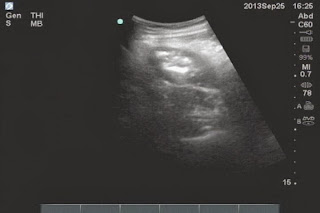
Ultrasound  abdomen first  showed  that  fluid collection around  liver and pelvis with one mass size of 3cm-4cm   at the painful area  (left lower abdomen)  like pseudokidney sign.


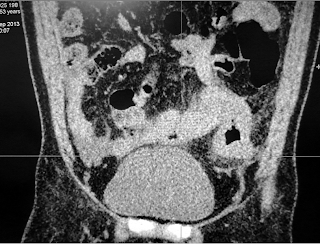
MSCT  without CE of abdomen detected  one mass with  intraluminal air and its wall was thickened more than 1cm which suggested inflammation like enteronecrosis.


This patient promptly was sent to BINH DAN HOSPITAL.and  abdomen x-ray for  check up  was done [see  photo].


Blood tests=   WBC rising 17K with  88% neutrophil.

Emergency operation performed as about peritonitis.due to perforation.
This mass is of  small intestine.which was looked like tumor or inflamation.


.

Microscopy reports that a cancer, But we have to wait for immunohistostainning to make sure that a malignant GIST or Carcinoid.

REFERENCE

Sunday 15 September 2013

CASE 209: A LUNG MASS, Dr PHAN THANH HẢI, MEDIC MEDICAL CENTER, HCMC, VIETNAM

Man 59yo in check-up lung x-ray detected a mass at left lung, asymptomatic, no history tuberculosis, non smocking patient.


Ultrasound of the lung showed that was a cystic avascular with well bordered mass, size of 4 cm.


MSCT CE said that cystic mass bilobar looked like a bronchogenic cyst.


Operation via endoscopic thoracotomy, and punction of this mass leaking out the milky fluid. Resection this tumor with macrospecimen picture.
Wait for pathology report, but the surgery report said it looked like a caseum cyst of tuberculosis.

REFERENCE: