Total Pageviews

Thursday 21 December 2017

CASE 468: ADRENAL GLAND TUMOR, Dr PHAN THANH HẢI, Dr NGUYỄN PHỨƠC TÒAN, Dr VĨNH TUẤN, MEDIC MEDICAL CENTER, HCMC, VIETNAM


Man 26yo,  ultrasound of abdomen for screening  incidentally detected  one mass of 5 cm at the right adrenal gland area.
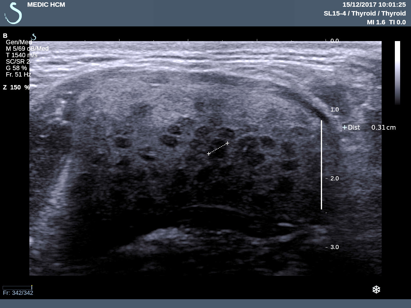
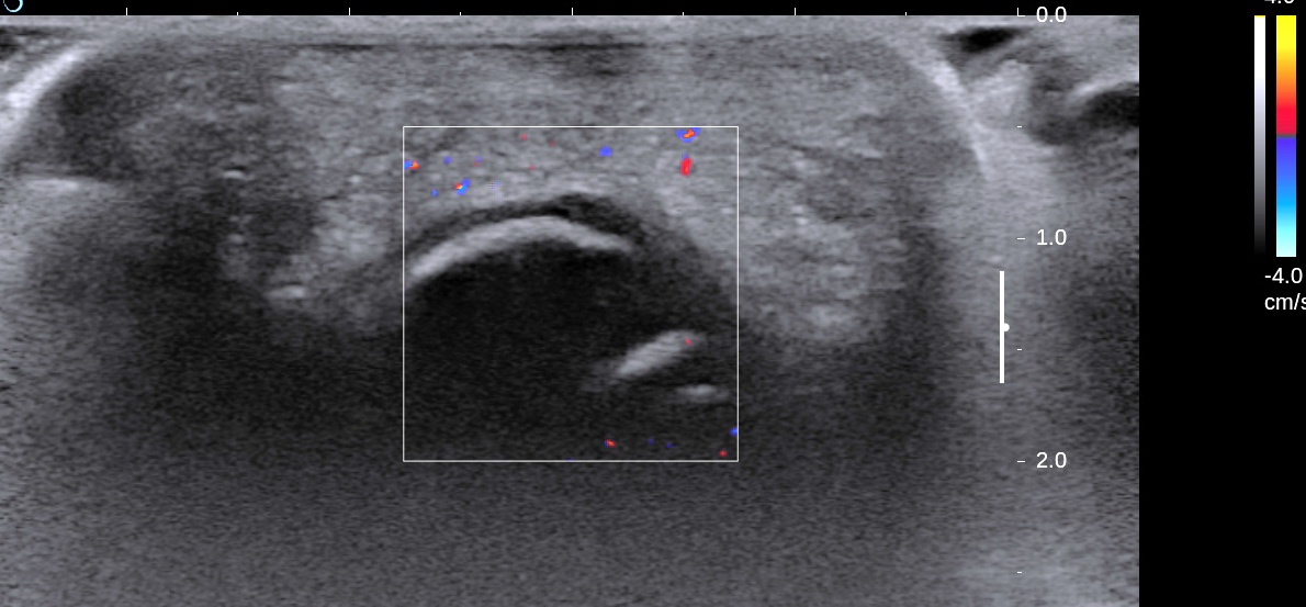
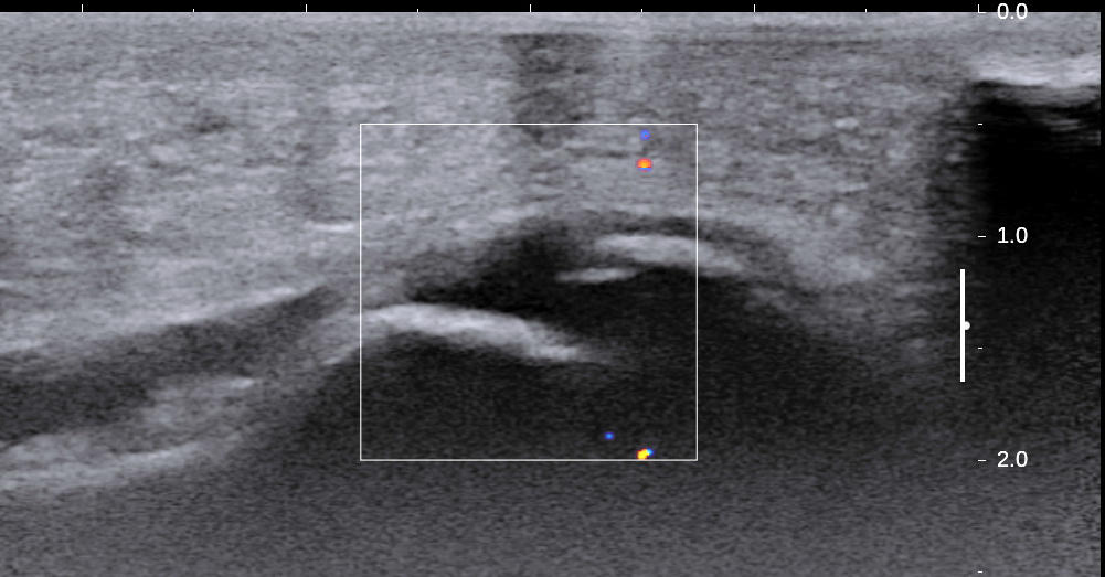
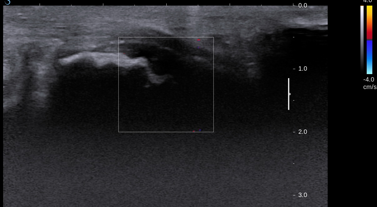
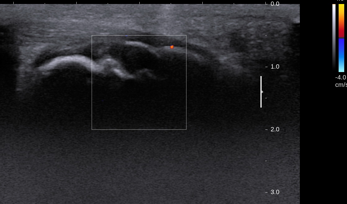
US1= longitudinal scanning of  this tumor at upper area of right kidney, well bordered.

  
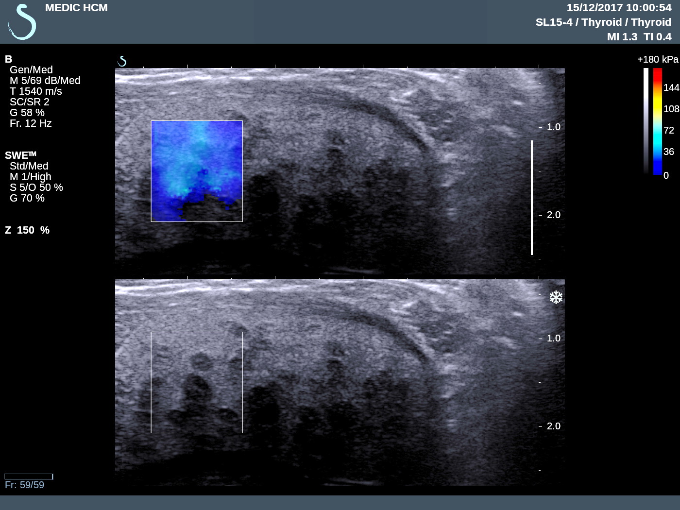
US2=  CDI of  this mass pulled down right kidney.


US3=  crossed section  of this mass is well bordered  under liver near IVC.


US4=  very  small vascular signals  in mass.


  • US5=   elastoscanning of this mass: very hard  32 kPa in comparison to  liver = 9.3 kPa.


Sonologist reported solid adrenal tumor for this mass.
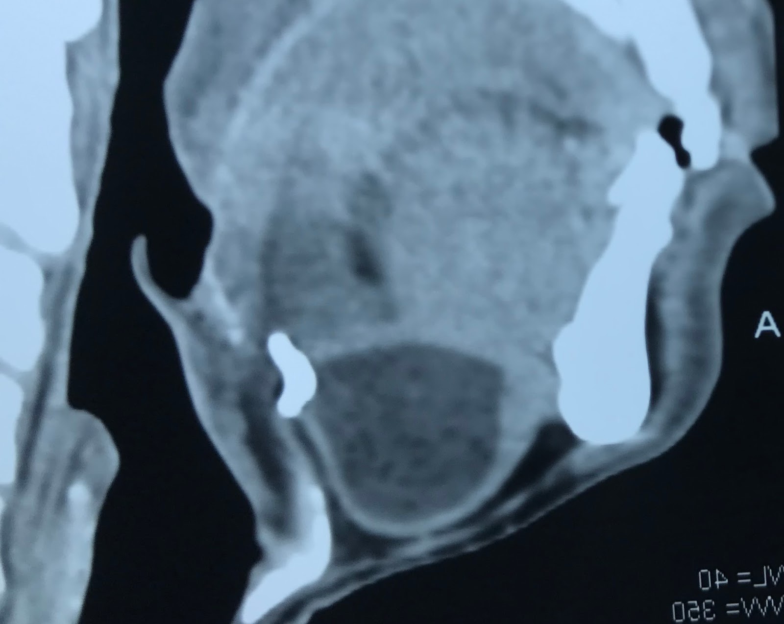
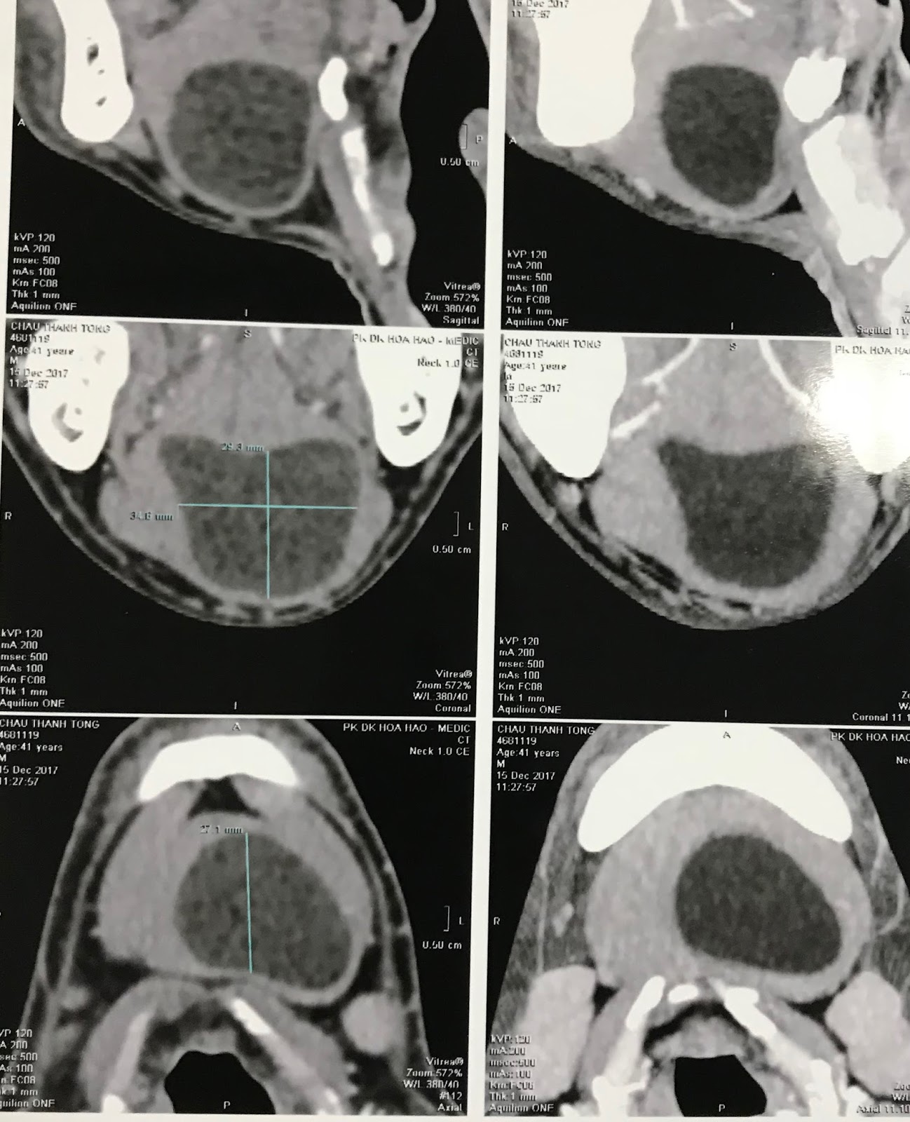
MSCT revealed:
CT 1= this mass is  hypodense like a cyst.


MSCT with CE, CT 2=  this mass is very low enhancement.
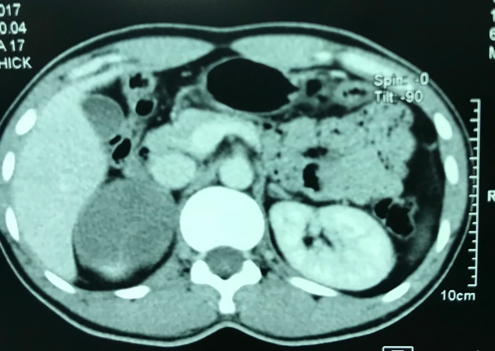

CT 3=  crossed section and sagittal scanning like a cyst of adrenal tumor.


Blood tests :  no abnornal  of cortico-medullary  adrenal function.
Pre-op   suggestion of surgeon is cyst of adrenal gland.
OPERATION REMOVED THIS TUMOR COMPLETLY.  MACROSCOPIC SPECIMEN WAS  WHITE AND HARD STRUCTURE, SECTION SURFACE SWELLED UP.
MICROSPIC REPORT IS  PARAGANGLION NEUROMA, BENIGN TUMOR.


REFERENCE:

https://journals.viamedica.pl/endokrynologia_polska/article/view/EP.2014.0017/32252


Tuesday 19 December 2017

CASE 467: SUBMANDIBULAR MASS, Dr PHẠM THỊ THANH XUÂN, Dr PHAN THANH HẢI, MEDIC MEDICAL CENTER, HCMC VIETNAM


Man 41 yo,   5 years ago  detected one mass at submandibular region, slow growth. Clinical palpation no pain,  soft, and without  trouble of  eating.


Ultrasound:
US 1=  longitudinal  scan with  curve probe 3.5 MHz :   ovoid mass clear border, hypoechoic with  no vascular signals inside.


US 2 = scanning with linear probe 12MHz=   inhomogeneous structure with many  black spots,    size # 0.5 cm.


US 3 =  elastoscanning of   this mass disclosed a cyst  with  many spots  hardening;  like pomegranate fruit.


CT scan with CE=   it is a cyst,  well bordered, .CT1, 2 , 3 with 3 sections of  this mass,    radiologist said  teratoma.


Operation for  remove this cyst with  content like  yellow milk   typical of  sebaceous cyst  (epidermoid cyst).


 MICROSCOPIC REPORT  IS EPIDERMOID CYST,   BENIGN TUMOR.


REFERENCE: IT IS RICE BODIES.


Sunday 17 December 2017

CASE 466:CHILDREN HEEL PAIN : A SEVER'S DISEASE CASE , Dr PHAN THANH HẢI PHƯỢNG, MEDIC MEDICAL CENTER, HMC, VIETNAM

9 years old male patient,  with chief complain of pain in both heels, which worsen by physical activities such as walking, running.
Physical examination: generally normal, Squeeze test (+) on right side.

 X-ray examination and ultrasound were performed.

On ultrasound plantar fascia is normal. Note: the anechoic region between calcaneous is not fluid (which can indirectly suggest fascilitis in case of adult) but in fact the normal apophysis (growth plate). 


Achilles tendon is normal and remains continous fibrous echotexture (US 2), again, the rough bone surface with anechoic shown normal apophysis.

Normal distance to apophysis in both sides, no dislocation, no avulsion.


X-rays examination of both 2 heel  are normal.


Physician suggests Sever's disease, and patient was told to take some time to
rest, proper physical activity and shoes fitting.

Conclusion:
Sever's disease, the most common cause of children heel pain, known as calcaneal apophysitis is an inflammation of growth plate in heel of growing children.  Diagnosis usually bases on clinical,  and X-rays is normal. Ultrasound is suitable diagnostic tool while X-ray examination is only helpful when an ossification center of apophysis exist. Ultrasound  helps ruling out muscle strain, detect edema, lytic and avulsion.