Total Pageviews

Wednesday 18 April 2018

CASE 488: CT SCANNING INCIDENTAL DIAGNOSIS of RECTUM CANCER, Dr PHAN THANH HẢI, MEDIC MEDICAL CENTER, HCMC, VIETNAM


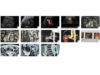
Woman 79 yo  being treated  right kidney stone.  3 years ago, with  ultrasound of abdomen in  black and white images   detected a stone with size 1 cm (US 1, US 2   longitudinal scan and crossed  section of right kidney).


On color Doppler  it exists an AVM  with calcification ( US 3, US 4).


MSCT with CE of abdomen  for   sure  AVM  of right kidney (CT 1,CT 2, CT 3, CT 4, CT 5) , radiologist reported at  CT 3 image  one mass  at  pelvis  like  sigmoid colon  tumor.


Coloendoscopy  confirmed that  rectum tumor  # 16 cm, high from anus.


Biopsy on the way (endoscopic image)
MRI  preop  takes staging of rectum cancer T4 N1 Mx.


Conclusion: Abdomen CT for  diagnosing AVM of right kidney detected incidentally a rectum cancer on AVM and stone kidney patient.

Operation  for  resection of rectum tumor and reanastomosis by stappler (MACRO ).


MACROSCOPIC  REPORT  IS  ADENOCARCINOMA  OF RECTUM.


SUMMARY  = CTA  IS THE BEST DIAGNOSING MODALITY for RENAL AVM  at THE SAME TIME  OF INCIDENTAL DIAGNOSING FOR RECTUM CANCER  WHICH WAS MISTAKED BY ULTRASOUND.
Monday 9 April 2018

CASE 487: BRAIN AVM, Dr PHAN THANH HẢI, MEDIC MEDICAL CENTER, HCMC, VIETNAM


Woman 39yo with headache and epileptic crisis.
TCD Ultrasound: TCD  1  from right temporal to left.
TCD 2: from left to right detected  Doppler vascular signals.
TCD transorbital  R/L   detection of left eye Doppler vascular with spectral pattern   TCD 4


MRI of brain=  MRI1: crossed section, mass vascular at  left brain.
MRI 2:  big  AVM  at left brain
MRI 3:   sagittal section, big vein  drainage from  AVM.
MRI 4 : sagittal  at left  brain..near left orbital area.
MRI 5:  MR angio:   AVM of left brain.


Wait for DSA  treatment.
Conclusion:  TCD ultrasound  of  the brain  detected left brain AVM.


Friday 6 April 2018

CASE 486: POLAND’S SYNDROME, Dr PHAN THANH HẢI, Dr TRẦM THỊ TÚ HƯƠNG, MEDIC MEDICAL CENTER, HCMC, VIETNAM


Woman  42 yo with  right breast hard mass,clinical examination of asymetric thorax (photo).


Ultrasound of right breast detected one 5 cm mass, multilobular, hypoechoic  with blood supply arround this tumor ( US 1, US 2 ),  US 3  detected axillary lymph node, US 4  in comparison of right to left chest wall  shows absence of right major and minor pectoralis muscles.


Chest X-Ray : clear right lung in comparison to left  lung due to  right chest wall muscle defect.


Mammography  diagnosis is  breast tumor with  Bi-Rads 4  T2N1Mx.


MRI   made diagnosis of  right breast tumor  with   pectoralis muscle defect of chest wall: it  is Poland’s syndrome.


FNAC of  this tumor  with cytological report of  adenocarcinoma of breast tumor.


Reference  : Case report   Poland ' s Syndrome complicated with breast cancer.

Tuesday 3 April 2018

CASE 485: CONGENITAL RENAL LYMPHANGIOMATOSIS, Dr PHAN THANH HẢI, MEDIC MEDICAL CENTER, HCMC, VIETNAM


Female patient 15 yo with  pain at lumbar region.
Ultrasound and CT detected  perirenalcystic  collection, kidney deplacement no stone no hydronephrosis. CT  urology with CE: no leaking of contrast to fluid,  but urologist suspected  urinoma  ( CT 1, CT 2,  crossed section , CT3  frontal section, CT 4  2 kidneys  deplacement, CT 5 urinary system).


Puncture of this fluid  analysis is not urine,  like  lymphatic fluid.
Operation at right kidney  with argon laser for ablation of right renal capsule, and the left kidney was removed perirenal capsule.
Ultrasound of 2 kidneys shows  normal structure with vascular supply  (US 1, US 2, US 3, US 4  ascites).


And  MRI   after 3 months of  operation=The right kid has  many perirenal cysts,    and  left  kid  has fluid  collection arround without hydronephrosis ( MRI 1, MRI 2)​​.


Conclusion:   this case  is a congenital renal lymphangiomatosis case.

REFERENCE: