Total Pageviews

Friday 16 February 2018

CASE 478: POPEYE’ SIGN of BICEPS MUSCLE, Dr PHAN THANH HẢI, MEDIC MEDICAL CENTER, HCMC, VIETNAM.


Man 58 yo playing tennis got pain in left upper arm.  Clinical  detected left biceps muscle protrusion  in contraction and painful. ( see photo 1 relaxed position,   photo 2  flexion position).


Ultrasound of biceps muscle  =

US 1 :  longitudinal scanning represented   rupture at the upper head of biceps muscle.


US 2 :  crossed section disclosed  echo poor pattern due to hematoma.  


US 3 : crossed section at middle part of biceps muscle  showed  hyperechoic pattern  by contraction.


MRI  made sure that rupture of upper part of biceps muscle.


Operation for repairing the ruptured muscle.   

Conclusion:  Orthopedic pathology due to sports named  Popeye' sign. 

Reference:   Anatomy of biceps tendon  and  Popeye’ sign. 


Tuesday 13 February 2018

CASE 477: LEFT EXOPTHALMIC EYE, Dr PHAN THANH HAI, MEDIC MEDICAL CENTER, HCMC, VIETNAM


Man 52 yo with  cough and pain at left eye, protrusion the orbis and edema the cornea for one week (photo).

MRI of the eye and or brain.


MRI 1=   frontal view  of the left exopthalmic eye.
MRI 2=  frontal section of the left orbis.  edema of the  intra orbis muscles,
MRI 3=   crossed section of left orbis  : left superior opthalmic vein dilated.  
MRI 4=   the muscles in orbis are edema  and  cavernous sinus  is not  abnormal.
For  make  sure diagnostic  DSA was done that  detected  A-V fistula at  cavernous sinus.
DSA  with dilated opthalmic vein=   DSA1  putting of the coil,   DSA 2 after treatment. Coil embolisation is  spectacular reduction clinical sign  (photo 2).


The left eye returns  near normal 24 hrs after treatment.


Conclusion:    Basis clinical signs of MRI and DSA   can make diagnosis  and spectacular  treatment. success. 

Reference:  Anatomy of eye circulation.


Friday 9 February 2018

CASE 476: ABDOMINAL WALL TUMOR, Dr PHAN THANH HẢI, Dr VÕ NGUYỄN THỤC QUYÊN, MEDIC MEDICAL CENTER, HCMC, VIETNAM.


Woman 60 yo in general check- up,  ultrasound of abdomen detected one mass  6x4 cm  at RLQA.

US 1:  longitudinal scanning over pelvic region  disclosed one mass near urinary bladder.

US 2:  crossed –section  of  hypoechoic mass,  well bordered,  no change position when patient moves  to lateral decubitus.


 US 3:  CDI = hypovascular mass. The vascular supply of tumor from the abdomen wall.


 US 4 : linear probe presented  the tumor and abdominal wall.


US 5:  elastoscanning of   inhomogeneous structure of tumor.

Blood test is normal
MRI  with gado: 3 position scannings of  this tumor . Radiologist says  mesenteric tumor.


Laparoscopic operation: This tumor is extra peritoneum.  Macro view looked like Desmoid tumor.


MICROSCOPIC  PATHOLOGY REPORT IS  DESMOID TUMOR.


REF  CASE  361  MEDIC


REFERENCE: CASE REPORT.


Thursday 1 February 2018

CASE 475: INCIDENTAL THYROID CARCINOMA ON CT, Dr PHAN THANH HẢI, MEDIC MEDICAL CENTER, HCMC, VIETNAM.


Man 55 yo for check- up  MSCT  total body and blood tests.

Radiologist report  detected one mass # 1.6 cm at right lobe  of thyroid gland,  with  HU 91 UI  in comparison to  the thyroid tissue background =121UI ( CT1  CT2  CT3).


Blood test is normal thyroid function.

Ultrasound scanning is second look:

US 1:  crossed-section  of the hypoechoic  focal  lesion, well-bordered #  1.5 cm.
US 2:  longitudinal scanning of  the mass is 1. 7 cm, hypoechoic pattern,  with 
US 3:  CDI = hypovascular  mass,  no lymph node in the neck.


This mass is  in TI-RADS 3, need  FNAC.
FNAC report suspected  papillary carcinoma ( PTC).


Operation is done for subtotal thyroidectomy (see macro 1,2).


POST OP  PATHOLOGY REPORT IT IS PTC [PAPILLARY THYROID CARCINOMA].


REFERENCE:


Friday 26 January 2018

CASE 474: LEFT LIVER LOBE TUMOR, Dr PHAN THANH HẢI, MEDIC MEDICAL CENTER, HCMC, VIETNAM


Man  52 yo  with HBV chronic hepatitis ,    follow up  every 6 months  and negative AFP, HBV -DNA . But  ultrasound   detected  a big tumor # 6 cm in diameter in left lobe of liver.
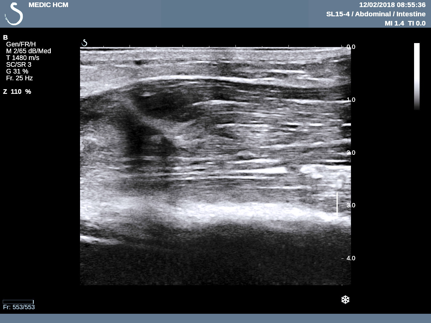
US  1:  transverse scanning of  this tumor is well bordered,   hypoechoic pattern.
US 2:  longitudinal scanning  at tumor site.
US 3:  color Doppler=  vascular supply to tumor is from left  liver.


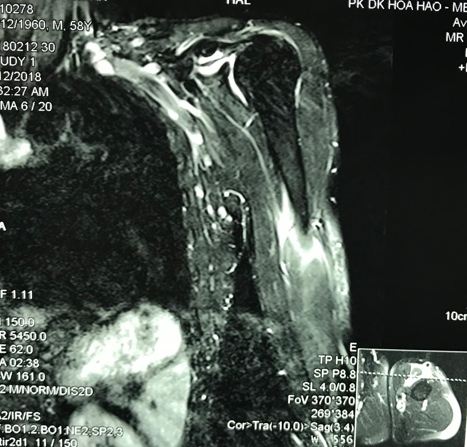
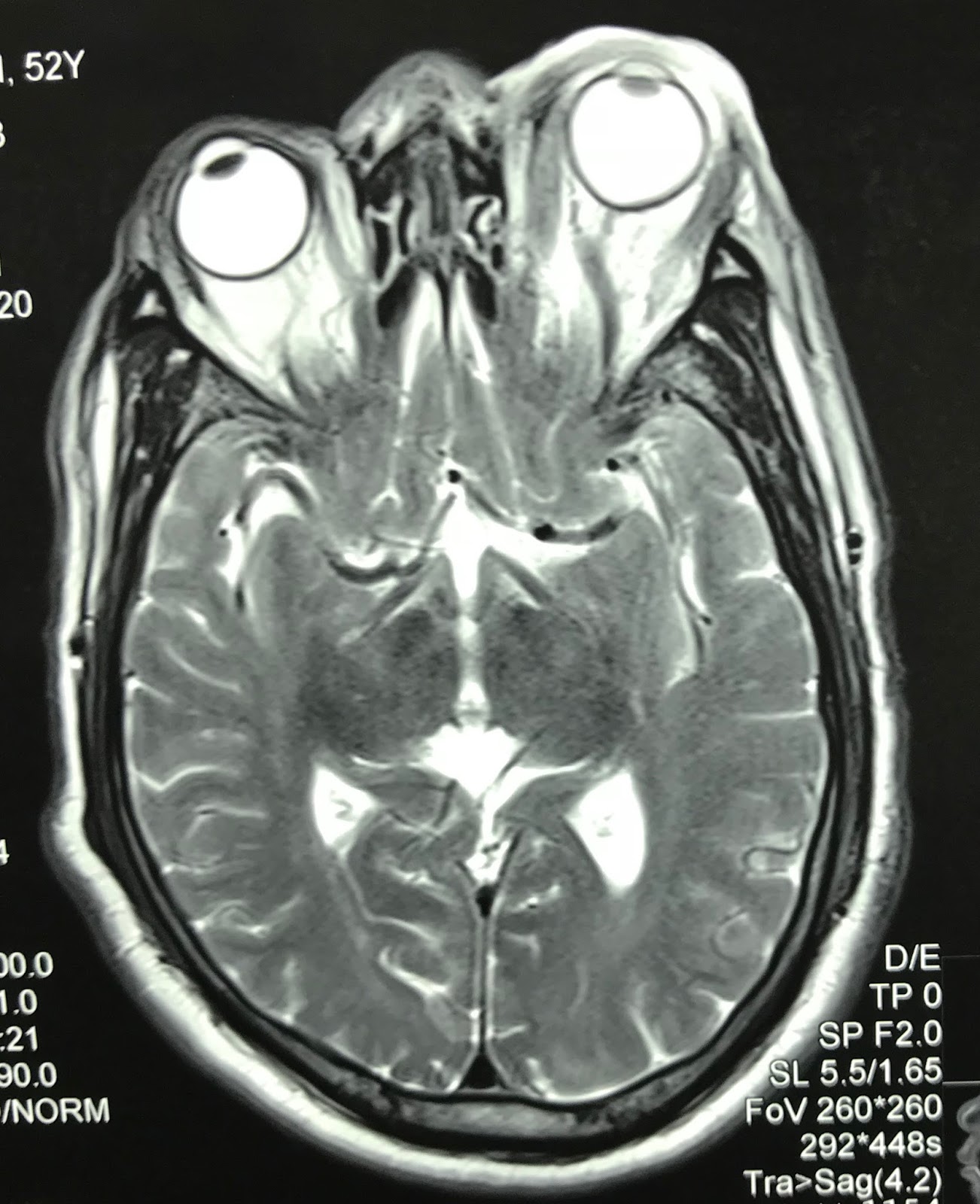
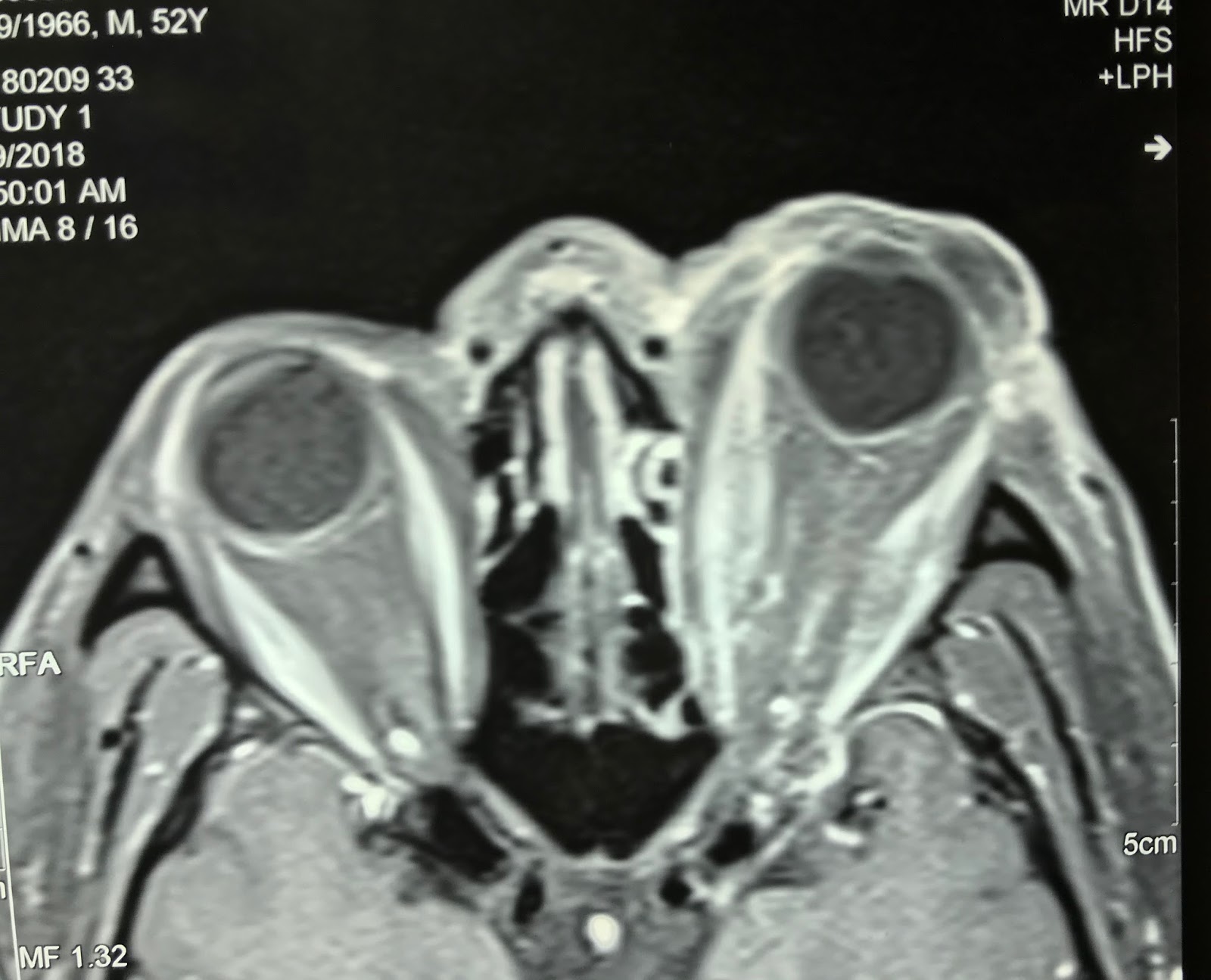
MRI  with Primovist  uptaked  and washed out  as a HCC in liver ( MRI 1, 2, 3, 4).


Blood test  Wako=  only DCP  raised =91 UI.


Summary:  in case of  HBV chronic hepatitis,, ultrasound detected  a big tumor in liver, Wako test only raised DCP.

OPERATION   PER OP  VIEW BY ENDOSCOPY THE TUMOR AT LEFT LOBE of LIVER ANTERIOR AND POSTERIOR and MACRO SPECIMEN of  TUMOR.


MICROSCOPIC IS WELL DIFFERENTIATED HCC.


DISCUSSION: WHY IN THIS CASE   WAKO TEST IS  NOT SUITABLE ?  WHICH VALOUR of WAKO TEST COULD BE  PPV FOR HCC ?

 Wako test post op  4 days after operation  = AFP:1.3 ng/mL;  L3: 0.5%;  DCP: 55 mAU/mL.

This 52 yo male  patient with chronic HBV but  AFP is lower than cut of value screening and no  ultrasound  screening  before  operation.   Wako test is  only DCP  rising  to 92 mAU/mL. Reference  ( publication April 12,2016 http:// doi.org/10.1371/journal.pone.0153227  : Diagnostic evaluation of DCP versus AFP  for Hepatitis Bvirus related  HCC   in China):

1-  30-40%  HCC in CLD with  AFP normal serum level.
2-  non HCC patients  have  15-58%  AFP  rise  over  cutt of value  20ng/mL.
3-  in CLD having cirrhosis  AFP rises  11-47%  but non HCC.
4-  DCP  rises level that correlated with size tumor and advanced progress.
5 - DCP drops very fast after surgery, and  rises  early in recurrent HCC.

Conclusion : AFP only not sensitive for screening  HCC. So DCP in Wako test is the choice for routine screening  HCC and monitoring after treatment.

BLOOD TEST  WAKO  AGAIN    RESULT IS  AFP: 1,5;   L3 : 0,5;   DCP DROP TO 38mUI/mL


CONCLUSION  2:

DCP IS VERY SENSITIVE FOR DETECTION of HCC AND FOLLOW UP POST OP.