Total Pageviews

Wednesday 4 October 2017

CASE 455: FINGER TUMOR, Dr PHAN THANH HAI, Dr TRUONG TRI HUU, MEDIC MEDICAL CENTER, HCMC, VIETNAM


Woman 54 yo,  4 months  ago  detected  one mass, at 4th finger of right hand,  slow growth,  no pain,  no disturbing movement.of this finger  (see  photo1, 2).


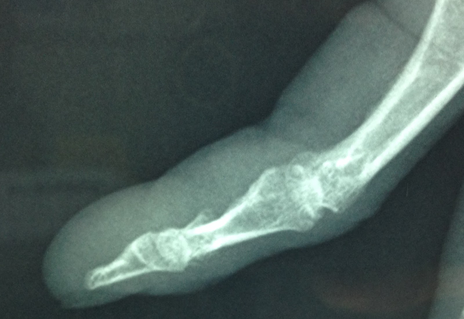
X-Rays  of AP and lateral views of 4th finger  = bone is  normal but  periosteum changing this mass to a soft tissue tumor ( xrays 1, 2).


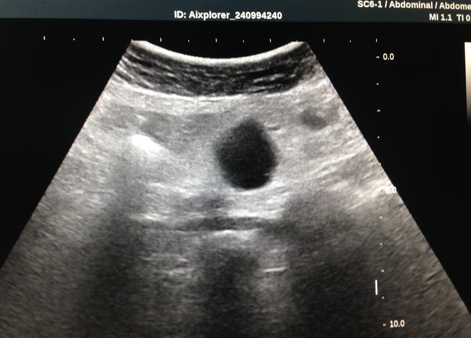
Ultrasound   scan of this mass is hypoechoic like a cyst of lateral finger, from the tendon,  size 3 cm of length (US 1).
US 2  CDI of  vascular supplying arround this tumor means  a solid tumor
US 3  crossed section  of the arround vascular tumor.
US 4 mass is soft  on elastoscanning , arround 30 kPa.


OPERATION REMOVED TOTAL TUMOR.

MACROSCOPIC  REPORT BY SURGEON LOOKED LIKE  XANTHOMA, BUT  MICROSCOPIC REPORT IS  GEANT CELL TUMOR of TENDON SHEATH.


Sunday 1 October 2017

CASE 454: IVC STENOSIS, Dr PHAN THANH HẢI, Dr TRỊNH DUY TRANG, MEDIC MEDICAL CENTER, HCMC, VIETNAM.


Woman 30 yo with 3 times of miscarriage, she came to MEDIC for a check- up (foto  subcutaneous veins).


Ultrasound of abdomen and pelvis:  normal uterus size.


US 1 = big liver caudate lobe 
US 2 = IVC  stenosis at  upper portion of liver
US 3 = crossed section of IVC no flow with hepatic vein.

MSCE with CE:


CT 1=  normal uterus structure.  CT2  = IVC  contrast filling  short portion cannot go upper to liver portion.  CT3 =  crossed section of  dilated subcutaneous abdominal veins. CT4 = crossed section= IVC no contrast in liver portion and abnormal late phase of liver vein, 
CT 5  = surface abdomen skin.


TREATMENT  ANGIOINTERVENTION;   DILATATION of STENOSIS AND STENTING.


Summary  =  IVC abnormal  stenosis near  diaphragm and  many venous collateral returning ways. 

REFERENCE:


Monday 25 September 2017

CASE 453: SMALL BOWEL MELANOMA, Dr PHAN THANH HẢI, Dr TRẦN MINH ĐÚNG, Dr DƯƠNG NGỌC THÀNH, MEDIC MEDICAL CENTER, TÂM TRÍ GENERAL HOSPITAL, HCMC, VIETNAM


Man 56 yo with acute abdomen pain,  vomitting, and dark stool [melaena]. Clinical examination was oriented to 4th day bowel occlusion.


Abdomen US scan in emergency  detected  dilated bowel  with  crossed sectional view  presented typical oignon sign  of intussusception  ( US 1,   crossed section;  US 2,  longitudinal  scan.    With linear probe, US 3, CDI examination;  US 4,  multilayer of  intussuscipiens [boudin].


MSCT  with CE of abdomen =   


CT 1: bowel dilatation  due to  bowel obstruction
CT 2 : mass  with  multilayer of small bowel wall.
CT 3 :  intussusception with target sign or pseudokidney sign
CT4 :  sagittal view of the abdomen
Lab test is normal.

Emergency  operation  via laparotomy  with diagnosis  intussusception by small bowel tumorSurgeon reported that  tumor is black color, intra jejunum, size 5 cm. Microscopic report  with immunohisto chemistry is  malignant melanoma.


UPDATE:

For DISCUSSION  whatever PRIMARY OR SECONDARY MENALOMA?
                       CAREFUL EXAMINATION FULL BODY  DETECTED  ONE SCAR AT THE  LEFT PLANTAR FOOT DUE TO OPERATION 6 YEARS BEFORE AT CANCER CENTER.
BUT PATIENT DID NOT REPORT THIS ISSUE and HAS NOT REPORT FROM THIS OPERATION.
THIS CASE  MAY BE CASE of SECONDARY MELANOMA METASTASIZING TO SMALL BOWEL ( SEE FOTO).


REFERENCE:   SMALL BOWEL MELANOMA
                 

Friday 15 September 2017

CASE 452: MESENTERIC CASTLEMAN DISEASE, Dr PHAN THANH HẢI, Dr NGUYỄN PHÚ HUU, MEDIC MEDICAL CENTER, HCMC, VIETNAM.

Woman 65 yo with  anorexia,  weigh loss # 5 kg in 3 months.
Ultrasound of abdomen for general check-up detected  one round mass, mobile  at  RLQ.

US 1 :  hypoechoic homogeneous  mass looked like a cyst,  size 3 cm, round border  with  low posterior enhancement,  intra mesenteric situation.


US 2 : CDI   vascular  around this mass not branching inside this mass   
US 3: CDI with another section, no vascular in the mass
US 4 scanning by linear 14 MHz  probe:  this mas  like a cyst.
US 5 :  elastoscan of this mass  mean  9 kPascal.

MSCT with CE=


CT 1:  crossed section of this mass very quick and  high contrast enhanced,  

CT 2:  frontal section of  this mass = intra mesentery

CT 3:  sagittal section of this mass:  near  anterior wall of abdomen  

Blood tests= normal  CBC, negative all cancer markers.

DISCUSSIONS=

 1. This mass is  intra mesenteric area not related with GI TRACT

 2. THIS MASS IS VERY  HYPOECHOIC AND HOMOGENEOUS  LOOKED LIKE A CYST  BUT IT IS SOLID MASS

 3. THIS MASS IS VERY QUICK AND HIGH CONTRAST ENHANCEMENT, HOMOGENEOUS STRUCTURE.


 4.  BLOOD TESTS   BETA 2  MICROGLOBULINE and FERRITIN , LDH NOT RISING


THE FIRST CHOICE OF  THIS MASS IS LYMPHOMA.


Laparoscopic operation  detected the mass intra mesenteric  jejunum; open surgery removed this mass out the abdomen cavity and web resection.

Microscopic report with  immunohisto-staining is  CASTLEMAN DISEASE.


REFERENCES: With 2 VMU Cases in the past.