Total Pageviews

Saturday 5 August 2017

CASE 445: LIVER PEDUNCULATED, Dr PHAN THANH HẢI, Dr NGUYỄN PHÚ HỮU, MEDIC MEDICAL CENTER, HCMC, VIETNAM

Woman 33 yo  with epigastric pain  has been treated as  gastritis.
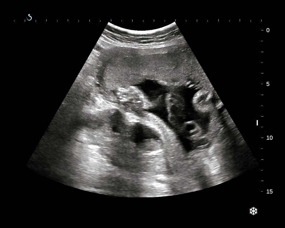
Ultrasound of  abdomen detected  one mass near  the liver border having hypoechoic peduncule from liver,  and changing position with  respiration movement (US1). There is vessel from the liver for peduncule of tumor (US ). In  cross-sectionnal scanning,  this tumor represented its well bordered, solid, hypervascular structure (US 3)


MSCT with CE detected this tumor in  connecting with  the liver by a long peduncule ( CT 1,sagital view) and in  frontal view, this mass is  nearby the  ligamentum falciformis (CT2).


CT 3: cross sectionnal scan of  tumor = well contrasted enhancement.
CT 4: vascular supplying for this tumor is a branch from  left gastric artery and  another one from liver.


Laparoscopic operation  detected  tumor  from  liver  at the border of falciform ligament,  and its structure  like liver tissue.


   Microscopic report  is  liver tissue.


Conclusion: it is a case of liver pedunculated .

Reference:


Sunday 23 July 2017

CASE 444: FEVER in PREGNANCY, Dr PHAN THANH HẢI, MEDIC MEDICAL CENTER, HCMC, VIETNAM


Woman 23 yo, 28 weeks primigravida with 5 days fever and abdominal pain in  periodic crisis.
WBC=  27 k  with neutro 97% ;  CRP= 342 mg/L,  normal urine test . And clinical suspected  appendicitis.
Ultrasound of  abdomen reported 29w pregnancy and  normal status of fetus. One cystic mass retro-uterus  near left pelvis  iliac artery, was suspected  left kidney  ectopic pelvis with hydro-pyonephrosis while  right kidney is normal ( US 1, US 2, US 3, US 4).


MRI  reported  left kidney hydronephrosis due to ureter obstruction.


Emergency cystoendoscopy was done for putting  stent  to release pus out from left kidney.


Conclusion=Pregnancy with  fever due to left pyohydronephrotic kidney as obstruction of ureter.

Reference :

http://www.bjuinternational.com/case-studies/hydronephrosis-pregnancy-ectopic-pelvic-kidney/


Monday 17 July 2017

CASE 443: DUODENAL TUMOR, Dr PHAN THANH HẢI, Dr LÊ ĐÌNH TÍN, Dr NGUYỄN PHÚ HỮU, MEDIC MEDICAL CENTER, HCMC, VIETNAM

Man 54yo with epigastric pain  crisis  after eating.
Ultrasound of abdomen  detected  tumor  at duodenum  D3, size of  3 cm,  hypoechoic (US 1),  and CDI  showed  hypervascular tumor  (US 2, US 3, US 4).


Gastrograph  with barium meal detected  filling defect  at  duodenum D 3 ( X rays1, X rays 2).


MSCT with CE:revealed  this tumor  very quick and high contrast enhanced (CT 1, 2, 3, 4).


Normal blood tests, and  nothing abnormal detected on endoscopy.

Operation for  resection of duodenum D 3  with the tumor from the wall  and central necrosis.


Microcsopic  this tumor is GIST.


Friday 14 July 2017

NHÂN CA 312 ĐƯỢC ĐĂNG TRÊN ULTRASOUND (UK) 2017, Vol 25(2)

CASE 312 : Fetal Body Hyperflexion { PENA-SHOKEIR phenotype} published on ULTRASOUND 2017, Vol 25(2) sagepub.co.uk/journal SAGE DOI: 10.1177/1742271X16688235

case 312 VUD

Xem fulltext theo link
https://app.luminpdf.com/viewer/ihfxeZZRFjjr8zsi3


Wednesday 12 July 2017

CASE 442: BILOBATE GALLBLADDER: Dr PHAN THANH HẢI - Dr LÊ THANH LIÊM, MEDIC MEDICAL CENTER, HCMC, VIETNAM. .


Male 18yo  with recurrent right subcostal pain.  
Ultrasound of abdomen detected abnormal gallbladder, size of 5 cm, bilobe, thickening wall of GB fundus  with more  twinkling artifact pattern on Doppler examination.


MRI of liver and biliary confirmed the morphology of gallbladder, its shape is bilobe and abnormal signal of GB fundus.


Radiologist report is gallbladder adenomyomatosis.


Operation laparoscopy removed the bilobate gallbladder.
Microscopic report is adenomyomatosis  of gallbladder.


Reference : US, CT , MRI diagnosis of   adenomyomatosis gall bladder