Man
51 yo with acute pain at right flank, type colicky
pain. History of being treated renal stone of left kidney by operation and ESWL for 2 years.
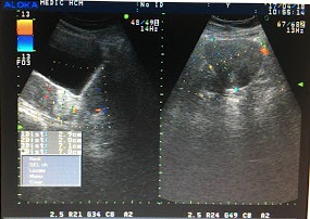
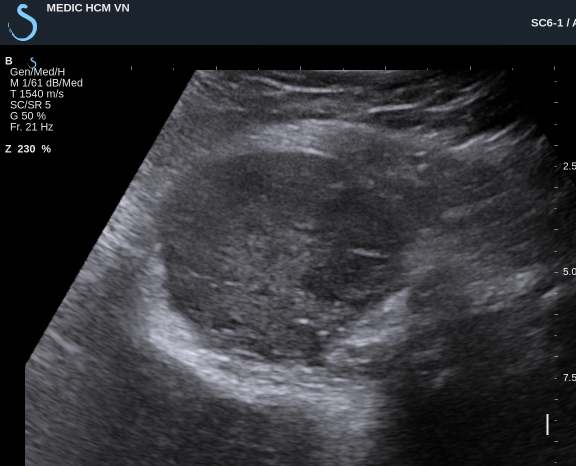
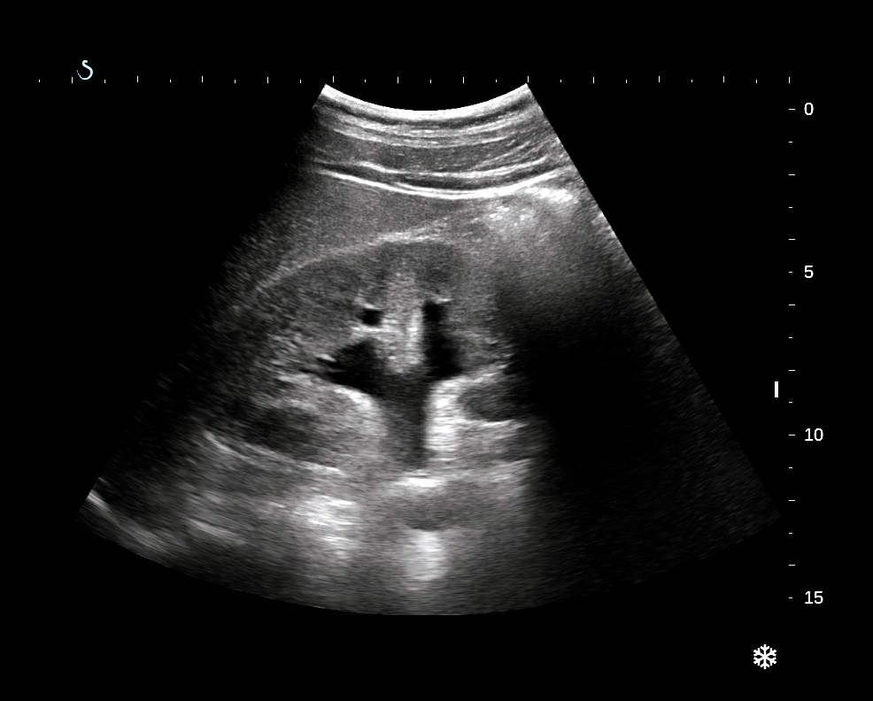
Emergency
ultrasound detected right and left kidney hydronephrosis (US 1, US
2).

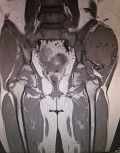
CT
scanning with CE: CT 1= kidneys no CE.

CT 2 with CE, arterial phase.

CT 3, venous phase.

CT 4, delay phase.

CT
5, frontal view.

CT 7, 3D view.

Blood
test: EGFR= 23mL/s.
Discussion:
Ultrasound scanning in acute renal colic crisis cannot make diagnosis of A K I ( acute kidney insufficiency); CT
non CE with HU low and CE phase in delay secretion
that suspected AKI.
Emergent
operation was done for removing of the stuck stone in right ureter for this
case.