Total Pageviews

Saturday 25 February 2017

CASE 418: CHEST PAIN in KAHLER DISEASE, Dr PHAN THANH HẢI, MEDIC MEDICAL CENTER, HCMC, VIETNAM


Man 59 yo with chest pain  at sternum.
Cardiac ultrasound  suspected  cardiac ischemia, but  EKG  is normal.


Blood  test in  emergency  report   troponin I HS is high 60ng/ml  
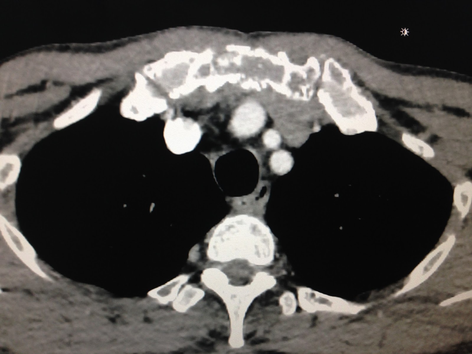
CT scan  cardio-thorax detected sternum is  erosion.


Blood test  again  after 2h troponin I HS is dropped to 53ng/ml   and troponin  T HS is 82ng/mL.,  PTH is  12ng/mL,  BETAMICROGLOBULINE 4254 ( high)    AND  KAPPA GLOBULINE detected in electrophoresis.

CONCLUSION   TROPONINE rise  ABNORMAL in KAHLER disease, not  due to cardiac  infarction..

REFERENCES= 2 TEXTs.


Monday 13 February 2017

CASE 417: BLACK SKIN TUMOR, Dr PHAN THANH HẢI, MEDIC MEDICAL CENTER, HCMC, VIETNAM


Woman 60yo with small skin  tumor at  righ antebrachial region which appeared 40 years ago, but now changing the color in black skin  without  pain, and hard in palpation.
(see  foto).


Ultrasound  of this tumor with 12 MHz linear probe.
 US 1: this tumor is underskin cover with  strong attenuation. Size of 1 cm.


US2 :  in CDI with very little small vascular signals.


US3: Changing of  angle of the probe, this tumor is  very high colored  like artifact.


US4:  In  this angle  twingling artefact appeared very strong.


Video  represented  twingling color of this tumor.


Operation with local anesthesia removed this mass that is metalic object.

Summary= It is  a foreign  body  made of metal  which was under skin for a long time made strong twingling Doppler artifact.

Reference:   Twinkling artifact on color doppler sonography.   AJR 2003,180:215-220.


Sunday 5 February 2017

CASE 416: RETROPERITONEUM HEMATOMA, Dr PHAN THANH HAI, VNT NHAN, NT TRANG,TC QUYEN, MEDIC MEDICAL CENTER, HCMC, VIETNAM.


Woman 56 yo with epigastric pain and anemia, RBC= 2.99m/L Hct= 28%
Normal colonoscopy.
US of  abdomen  detected one retroperitoneum mass near the head of pancreas, looked like hematoma ( US 1, US 2 ,US 3).


MSCT with CE confirmed  this mass is  retroperitoneum hematoma, maybe due to  rupture from a small aneurism of  gastroduodenal artery (CT1, CT 2, CT 3 CT 4  3D reconstruction).


Emergency  DSA  detected  the abnormal.variation of  hepatic artery and  small aneurysm of  gastroduodenal artery had stopped bleeding.


Survey this case for  Hct dropped  to 25% and  operation for control bleeding by  ligation this small ruptured aneurysma . 

Post-Op is in  well recovery . 


Conclusion: Easily diagnosed ruptured retroperitoneum aneurysm  by  combining of ultrasound, CT and, DSA.

Thursday 2 February 2017

CASE 415 : BREAST TUMOR, Dr PHAN THANH HẢI, MEDIC MEDICAL CENTER, HCMC, VIETNAM


Woman 57 yo, herselft  detected one mass at central left breast.
US 1: hypoechoic mass with size of  2cm at central left breast and one small nodule nearby which is sister tumor.


US2: CDI, this mass is hypovascular.


US3: Doppler PW of supply artery : PSV = 23cm/s  s/d=2.7. RI=0.62.


US4: ElastoUS, this mass is very hard. No axillary node detectable.


Mammo xray: this mass is no calcification, spiculated, classification BI-RADS 4.


Biopsy core reported this tumor is very hard.
Microscopic report with  histo immuno staining is  ILBC ( invasive lobular breast carcinoma). T2NoMx.


Summary: It is typical a breast cancer ILBC.


Friday 20 January 2017

CASE 414 : PANCREAS S P E N, Dr PHAN THANH HẢI, Dr PHÓ ĐỨC MẪN, MEDIC MEDICAL CENTER, HCMC, VIETNAM.


15 yo girl with epigastric pain.
Ultrasound  detected one 7cm mass, solid and cystic mixed echo at  tail of pancreas (US 1,2).


MSCT  with CE of this mass represented  round mass at the tail of pancreas, CE low enhanced ( CT1, CT2).


MRI with gado  (MRI 1, MRI 2, MRI 3, frontal view).


Radiologist reported  that solid  tumor of pancreas in suspecting of malignancy.
Blood tests: CA19-9 is low level, and other cancer markers are normal.
Operation (foto)  This round  border mass  pulled stomach and no evidence of spreading intra abdomen cavity.


Frozen section the mass and  microscopic report is  SPEN (solidary pseudo papillary  epithelial neoplasia of pancreas).


Conclusion: SPEN of pancreas.
REFERENCE:

http://casereports.bmj.com/content/2016/bcr-2016-218403.full

Friday 13 January 2017

CASE 413: INFANTILE HEMANGIOENDOTHELIAL of LIVER, Dr PHAN THANH HAI, Dr PHAM THI NGOC TUYET, Dr LE ANH TUAN, Dr LE QUANG THONG, MEDIC MEDICAL CENTER, HCMC, VIETNAM.


In  2008, a 5 months old  female baby with  abdomen distention and ultrasound scanning of her abdomen  detected  multiple echo poor  in her great liver ( US 1).


MRI of  liver  with gado  found out  multiple  white spots   with central  low enhancement, looked  like jack fruit appearance ( MRI 1, MRI 2).


CT of  liver non CE  also  found out multiple  black spot  inside her big liver ( CT1).


Blood test   with AFP = 76ng/mL.
Radiologist and hepatologist suggested that infantile hemangioendothelioma of liver,  no need of special treatment.
Reviewing of  this  case in  Jan  2017, now she is 9 year-old in well status, and  liver ultrasound  finds out only some small calcifications in her liver.


Conclusion: Multiple focal lesions of liver hemangioendothelial at neonate period now spontaneous regression for 9 years follow-up.


Update case 413 from Dr NGUYỄN ĐỨC TRÍ, CHILDREN N0 2 HOSPITAL


Today 18 Jan 2017, I see  an another case  looked  like the case 413 which is on a male 4 months old baby with abdomen distention.


Ultrasound and CT with CE  find out many round spots like jack fruit appearance.


Suspection of  a second case of liver infantile multiple hemangioendothelioma.

Wait and see  follow-up this case but how long to make sure the diagnosis?


REFERENCE: