Total Pageviews

Sunday 6 November 2016

CASE 404: ECTOPIC TESTIS TUMOR, Dr PHAN THANH HẢI- Dr NGUYỄN PHÚ HỮU, MEDIC MEDICAL CENTER, HCMC, VIETNAM.


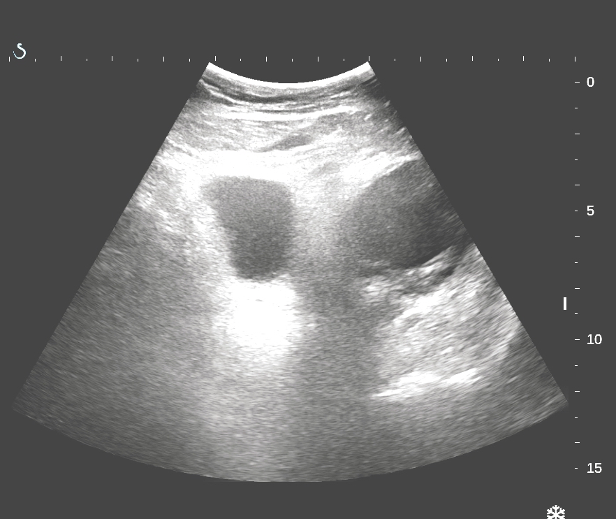
Man 35 yo with  pain at RLQ. Ultrasound  of abdomen 
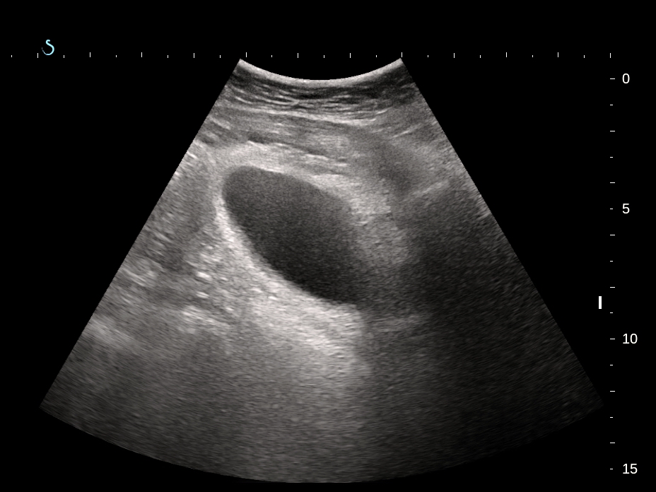
detected one hypoechoic mass retrocoecum ( US 1) which was  suspected an abscess.


Blood tests =  normal  
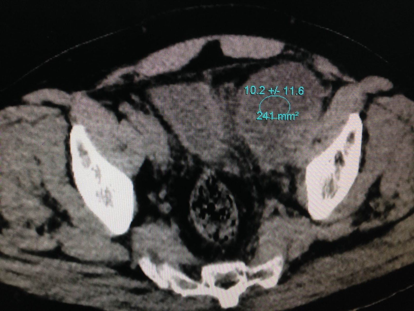
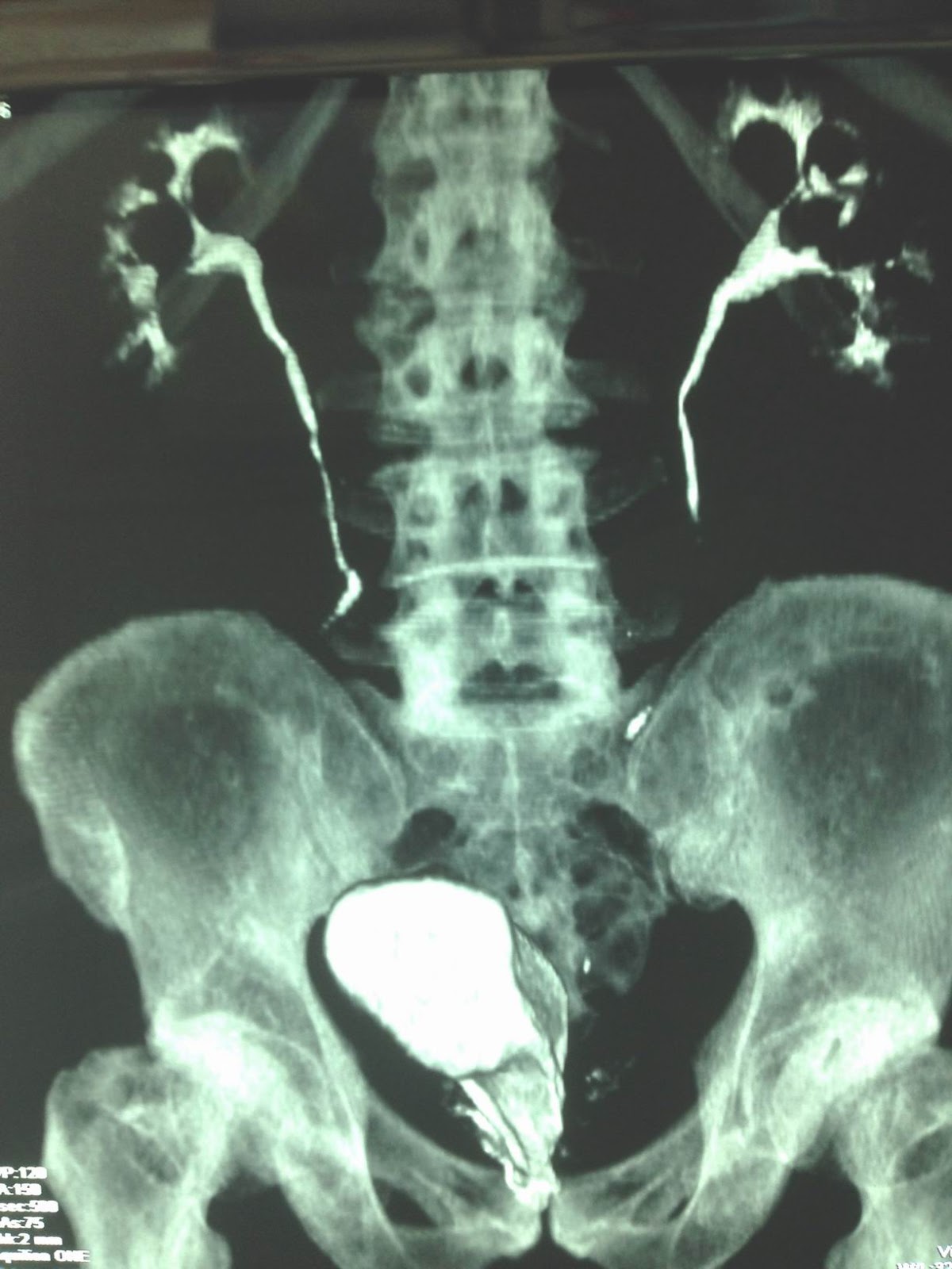
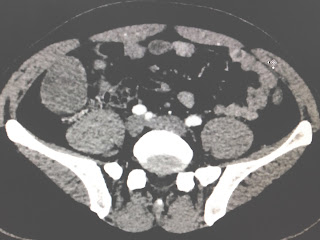
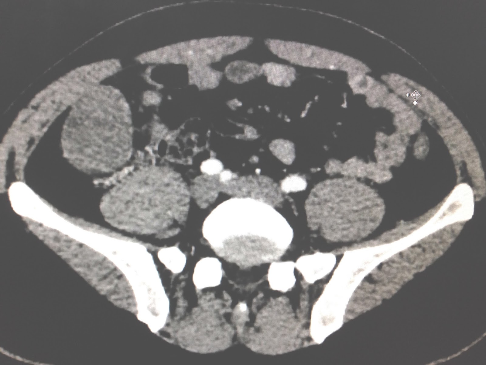
WBC and CRP. MSCT reported  this ellypsoid  mass with   size of 5 cm,   retroperitoneum, pull up the coecum, 
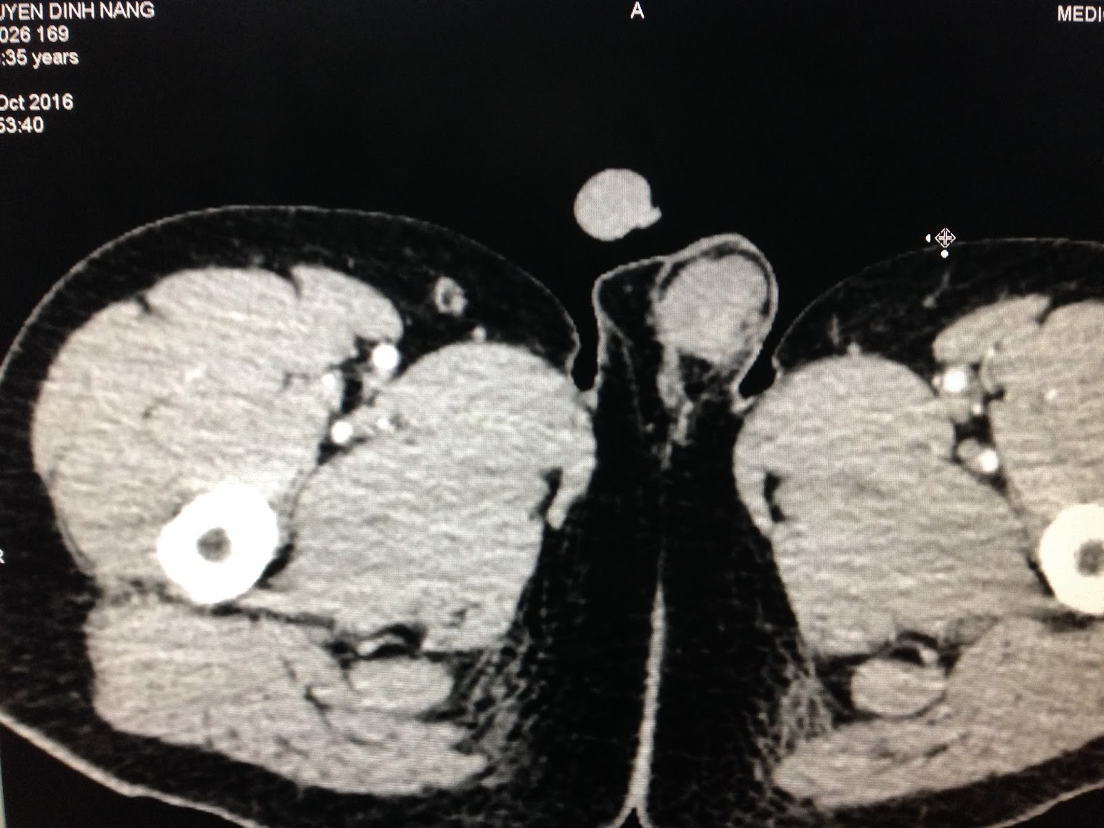
and MSCT with CE for rule out an abscess. (CT1: cross section, CT 2 sagital. CT 3  scrotum  section  not detected right testis.


Pre operative  surgeon suggested right  ectopic testis tumor.
Endolaparoperation detected  the coecum  was pulled  up ( ope 1, 2 ).


Ope 3=tumor is retro peritoneum.


Macroscopic removing this tumor which is  ectopic testis.

Microscopic  report is seminoma.

COMMENT: For  men  once ultrasound detected a hypoechoic mass in  retroperitoneum  at pelvis that has to verify ectopic testis, and if it is hypoechoic  like the cyst, the nature of testis tumor may belong lymphoma or seminoma.

Tuesday 1 November 2016

CASE 403: ECTOPIC ACCESSORY PANCREAS, Dr PHAN THANH HẢI - Dr LÊ VĂN TÀI, MEDIC MEDICAL CENTER, HCMC, VIETNAM.


Female 19yo with headache and  abdominal peristaltic pain. In check- up of abdomen,  ultrasound detected one  subhepatic mass  moving  with respiration,  size of 6 cm,  hypoechoic, hypervascular, not  attached to any organ in abdomen ( US 1, US 2) .


Upon  MSCT with CE, this mass  is  very fast  CE enhanced,  same time with pancreas ( CT1, CT2).


Blood tests are normal. And gastro-colonoscopy is normal.


Discussion:  CT and ultrasound showed  this tumor  in abdomen near the liver border and duodenum while scanning  in  standing position  this mass moving to right iliac fossa. One surgeon said  may be GIST.
Endolaparoscopic operarion  detected this mass is covered by great omentum (see foto 1) with very long pedicule due to great vascular supply (foto 2).
Removed this mass and  macroscopic this tissue looked like pancreas tissue (foto 3, 4).


Microscopic report  is  adenoma  of pancreas.


Friday 28 October 2016

CASE 402 : DOUBLE URINARY BLADDER, Dr PHAN THANH HẢI, MEDIC MEDICAL CENTER, HCMC, VIETNAM


Man 62 yo, one month after robotic prostatectomy by prostatic carcinoma (PC)  stage I, pain in mictation.
US scan at pubis detected 2 cystic masses  look like  double urinary bladder (US 1, transverse scan ).


US 2:  scan at  left  cystic mass ,  size of 4 cm, thickening of  border and septation.


US 3:  longitudinal  scan at  central cystic mass,  it is  urinary bladder.


US 4: left pelvis mass and  iliac artery.


Report of sonologist  is double urinary bladder.
MSCT with CE of  urinary system= CT 1, CT 2 non CE showed  HU units  at  2 masses are  different  15.8   and 10.2 UI.


CT 3 =  contrast filling of  the central  mass is urinary bladder and  left mass is not enhanced.


CT4 : 3D C T of urinary system.


Puncture of this cyst  for removing fluid which is yellowish. Results of analysis of  its contence : urea 
4.8mmol/L,
creatinine 0.0741mmol/L.
 So it is lymphatic fluid. Now  operation for drainage.
Conclusion:  it is lymphocele  post prostatectomy and lymphoadenectomy.

Friday 21 October 2016

CASE 401: BIG MASS beside HEART, Dr PHAN THANH HẢI, Dr NGUYỄN TUẤN VŨ, MEDIC MEDICAL CENTER, HCMC, VIETNAM


Man 41 yo with short breathing  in hard working. Chest X-Ray detected big mass in left upper lung (chest x-ray film).


Echocardio scanning  by cardiologist detected big cystic mediastinum tumor,  with think wall and calcification, size of 12 cm.


MSCT CE of thorax  and radiologist report is cystic teratoma.


OPERATION THORACOTOMY= THIS TUMOR IS  RUPTURED AND LEAKOUT  WHITE FLUID AND INTRA TUMOR IS SEBUM  AND SOME HAIR.


Microscopic report is mature cystic teratoma of thymus.


Wednesday 19 October 2016

CASE 400: NEONATE TESTIS TORSION, Dr PHAN THANH HẢI, Dr LÊ VĂN TÀI, MEDIC MEDICAL CENTER, HCMC, VIETNAM


Neonate  3 days old with  right swelling scrotum and left darker scrotum ( foto).


Ultrasound   US 1 = small right  testis  and  hydrocele vaginalis.


US 2 = left testis is  hypoechoic and no blood supply.


US 3 = cross section of  left testis.   


Sonologist reported  torsion of left testis  long time ago, maybe intrapartum stage
Operation= removed  left testis  torsion in 3 wings roller and black color.


Microscopic report is left  testis necrosis.


Conclusion=   Torsion of testis detected  at neonate time.

Reference:
http://www.jpss.eu/index.php/current-issue/item/558-bilateral-testicular-torsion-in-a-neonate-a-case-report


http://synapse.koreamed.org/Synapse/Data/PDFData/1020KJU/kju-49-957.pdf


http://file.scirp.org/pdf/CRCM_2013032513030136.pdf


Monday 17 October 2016

CASE 399: HCC GOING TO HEART, Dr PHAN THANH HẢI- Dr NGUYỄN TUẤN VŨ, MEDIC MEDICAL CENTER, HCMC, VIETNAM


MAN 66 yo with CHEST PAIN and DYSPNEA. EMERGENCY ECHOCARDIOGRAPHY DETECTED  INTRA CARDIAC MASS, LOOKED LIKE THROMBUS.
ULTRASOUND SCAN of LIVER DETECTED a LIVER MASS SIZE of 4cm and DILATED 2cm in DIAMETER and OBSTRUCTED by A HYPOECHOIC MASS ( US 1, US 2).


MSCT with CE DETECTED LIVER MASS and TUMOR INVASION TO HEPATIC VEIN TO IVC and GOING to RIGHT ATRIUM and FILLING DEFECTED at PULMONARY ARTERY (CT1, CT2).


CT3 (section) SHOWED LIVER MASS, RIGHT ATRIUM MASS and INTRA LEFT VENTRICULAR MASS.


BLOOD TEST = HCV POSITIVE; WAKO TEST = TRIPLE POSITIVE.