Total Pageviews

Sunday 13 March 2016

CASE 367 : INTRAORAL TUMOR, Dr PHAN THANH HẢI, MEDIC MEDICAL CENTER, HCMC VIETNAM


Boy  04 yo, difficulty  swallowing for 3 months.  No fever, no pain. Clinical  ENT  doctor’s  examination  is suggestion of tonsil  tumor at right side  (photo).


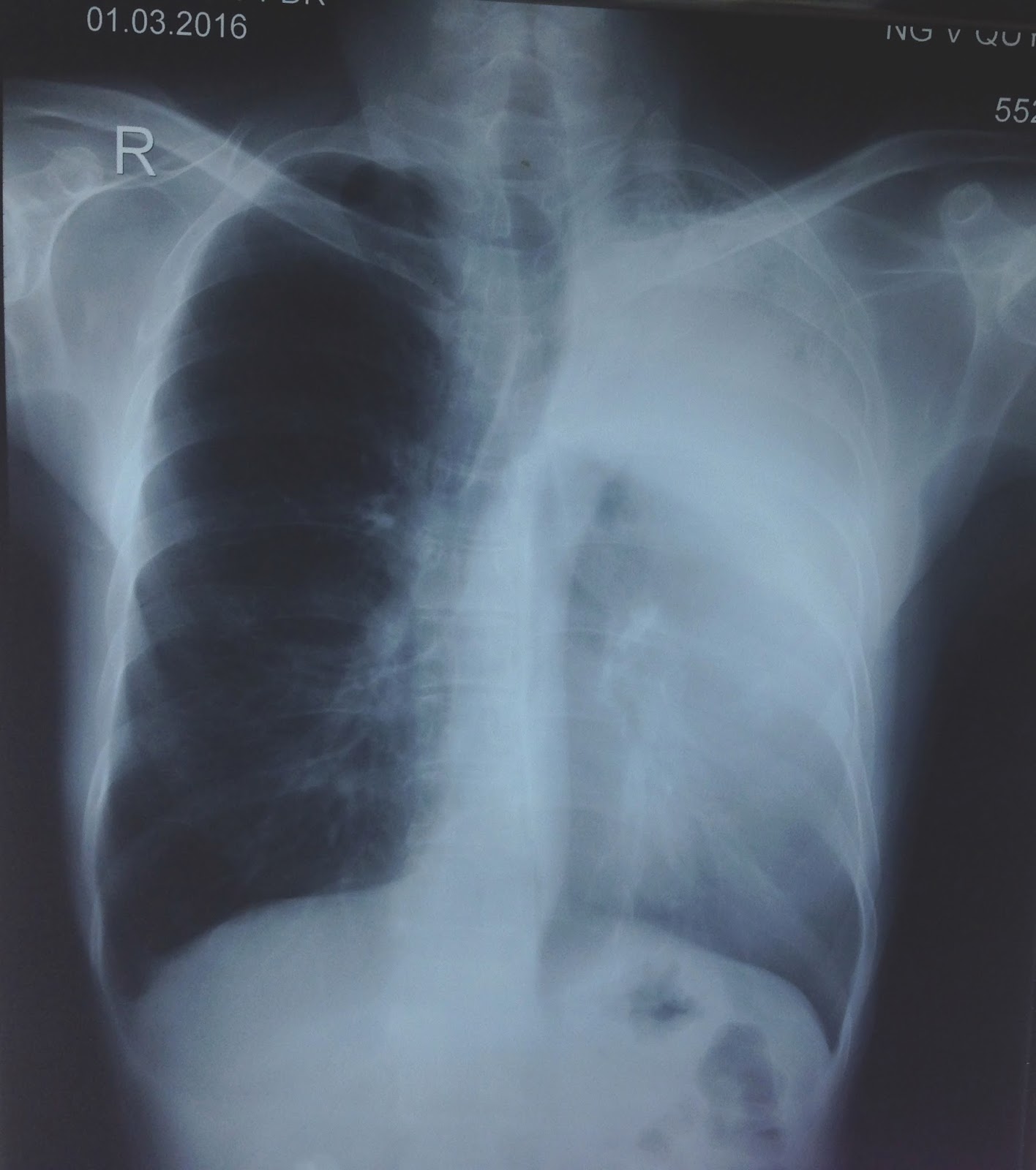
X-Rays of  the neck AP and lateral view: this mass  is  calcified,   irregular border,   precervical spinal bone, size of  4 cm ( film 1, 2)


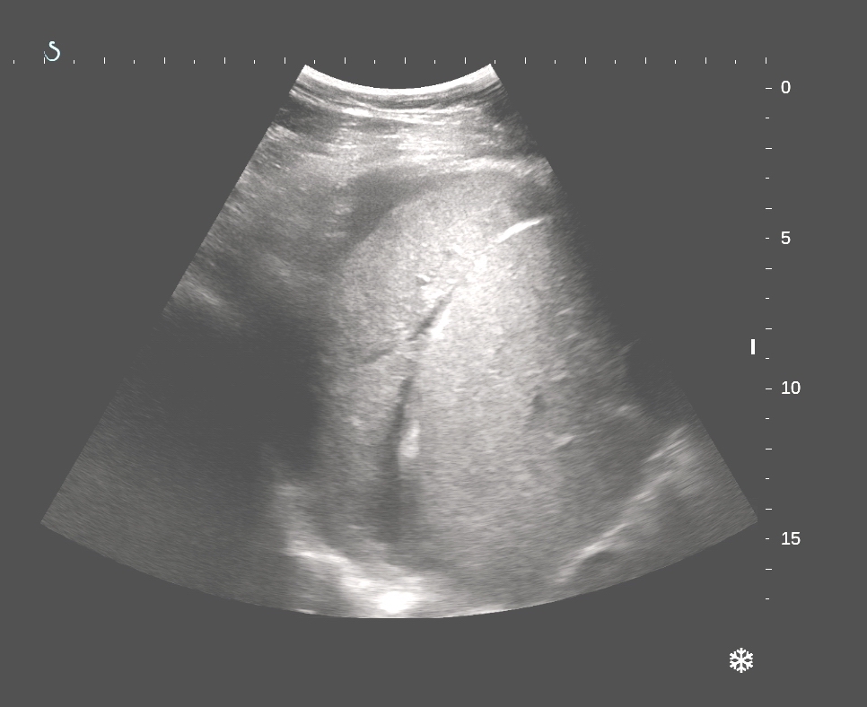
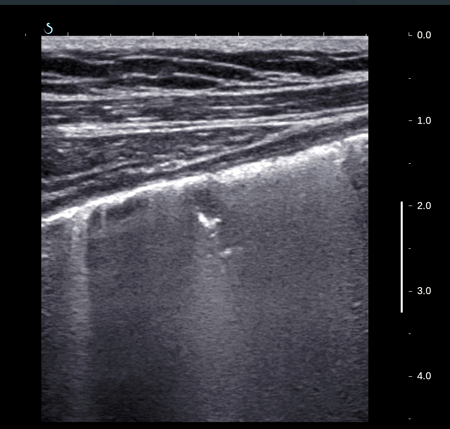
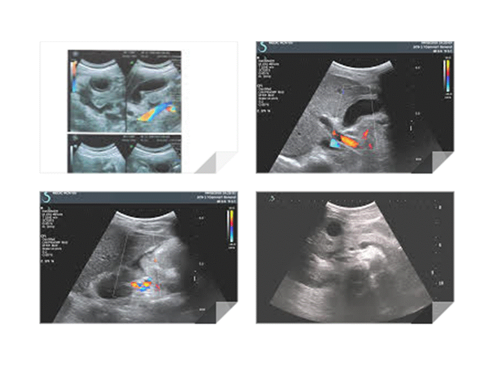
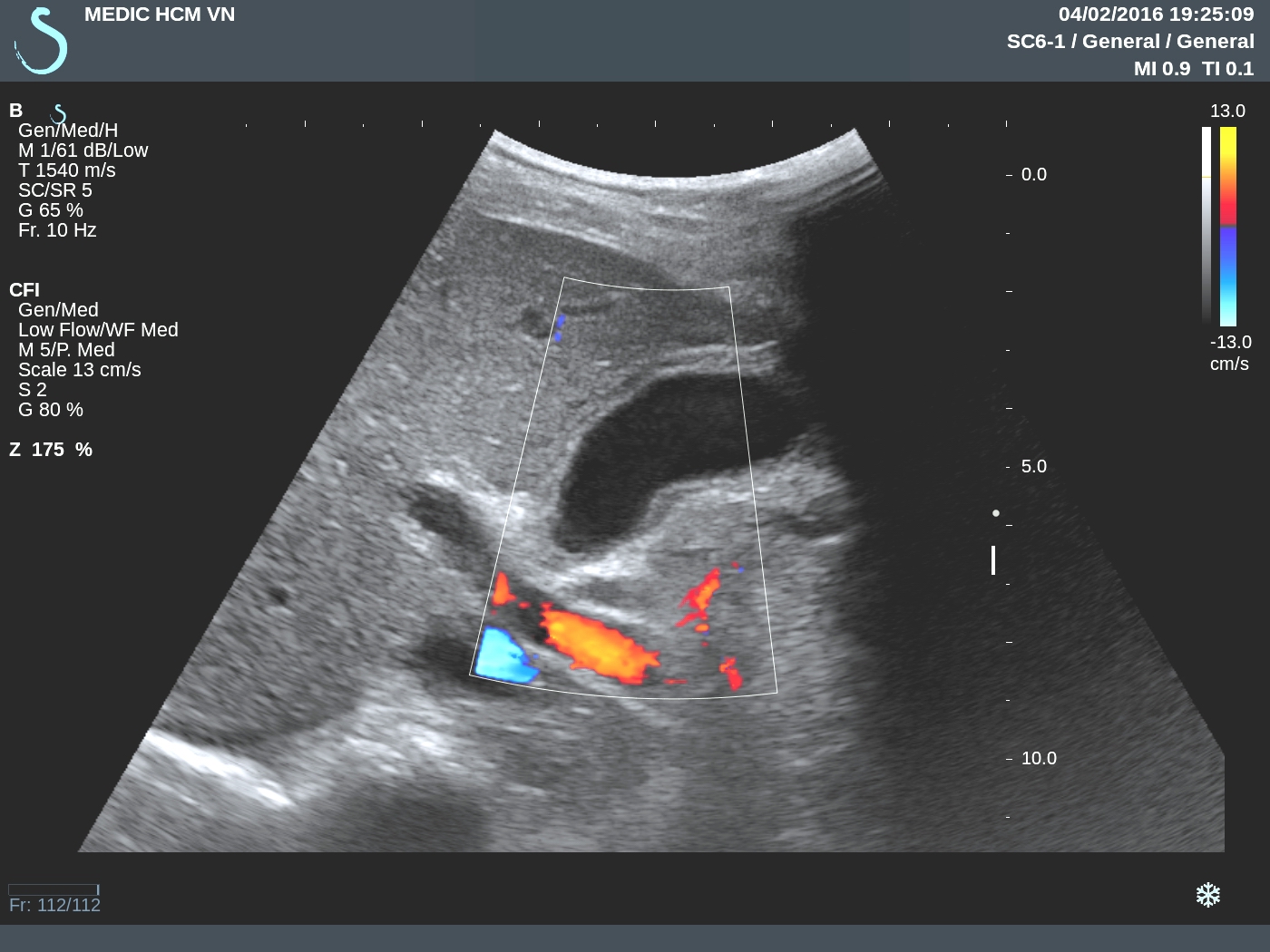
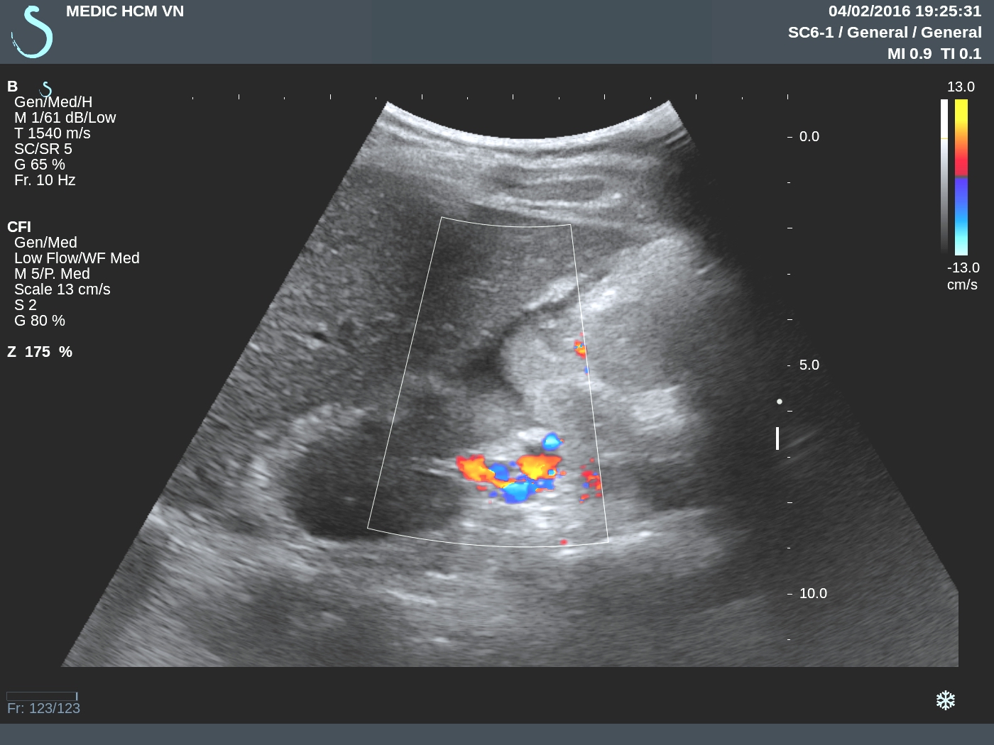
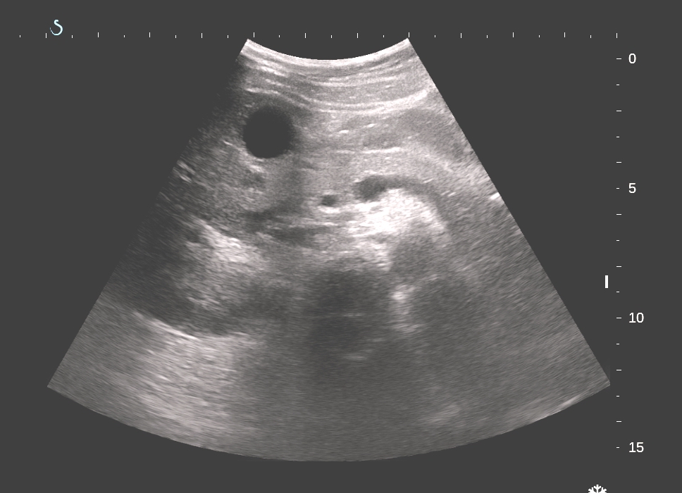
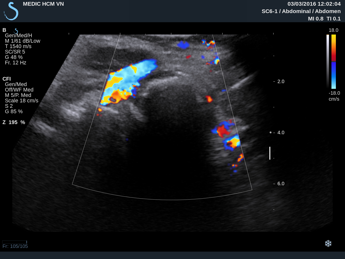
US examination of this mass:
US 1: Longitudinal scan  the mass with  strong shadowing cannot  inside this mass.

  
US 2: Cross-section view.


US 3: Relation of this mass with carotid and  cervical spinal bone.


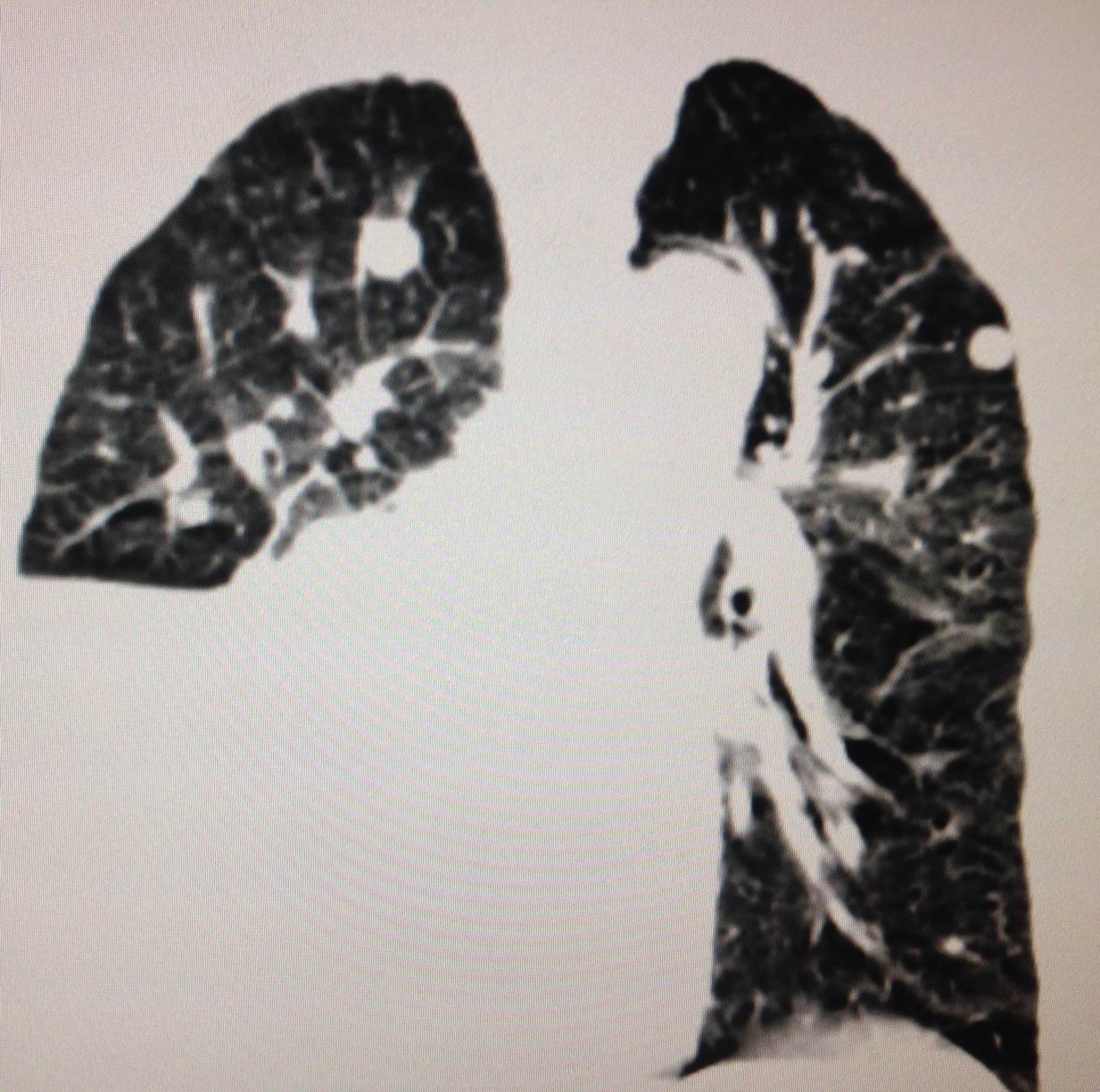
CT scan= CT 1:sagittal view , CT 2:  cross- section  with PA view,  CT3: cross-section with AP view.


Based on clinical, X-Rays , ultrasound and CT, what is your diagnosis?
Based on  X-Rays and CT  some doctors  suggested  teratoma of oropharynx, or enchondroma.
MRI ( 2 pictures sagittal and  section) radiologist  diagnosis is  chondroma.


Operation today  removed one  hard mass   looked like  stone.
Microscopic  report of this mass is fibrous dysplasia ossificans progressiva which is  same as  myositis ossificans


REFERENCE :  case  report.


Tuesday 1 March 2016

CASE 366 : LUNG HEPATIZATION, Dr PHAN THANH HẢI, MEDIC MEDICAL CENTER, HCMC, VIETNAM


Man 60 yo, cough, dypsnea.
Chest X-rays:(picture: very  bright left lung).


Ultrasound  scanning position on the back (photo)
U S 1:  scanning of the upper  portion of  left  lung, air  inside solid mass.


U S 2: lower portion of the mass=  echostructure  looked like structure of liver.


U S 3: vascularity of this mass.


U S 4 :small mass arround in  lobar bronche  is  consolidation of the lung, no pleural effusion.


CT 1  scanning  with  CE  detected  the mass  in left upper lobar bronche enhanced with CE.


Conclusion: Lung  consolidation on ultrasound  looked like  liver (hepatization of the lung)


Thursday 25 February 2016

CASE 365: MULTIPLE INTRAMUSCULAR TUMORS, Dr PHAN THANH HẢI, MEDIC MEDICAL CENTER, HCMC, VIETNAM.


Woman  60 yo being treated   lymphoma large B cell  stage IV by chemotherapy for 5 months.

One week ago she herself detected  many  subcutaneous nodules  palpable  at  forearm right and left, neck and  right parotid area, no painful.
 ULTRASOUND=
US 1=tumor  intramuscular right  forearm, round  border, very  low echo density.


US 2=cross-section, lesion at forearm.

US 3=CDI  Doppler vascular  structure of this mass, hypervascular.


US 4=longitudinal scanning   with  CDI.


US 5=CDI with PW,   RI = 0,70.


US 6 = small intramuscular nodule  at posterior of  neck.


US 7= SWE of mass in right  parotid.


Do you thing  it is lymphoma  in muscle?  
Biopsy of this mass  is large  B cell lymphoma, same as  result pre-treatment.


Conclusion: LYMPHOMA  LARGE B CELL  AT THE DIFFUSE STAGE  CAN MAKE  MULTIPLE NODULES  IN MUSCLES.
Reference:

Monday 15 February 2016

CASE 364: LUNG LOOKED LIKE LIVER, Dr PHAN THANH HẢI, MEDIC MEDICAL CENTER, HCMC, VIETNAM


Woman 62 yo, cough and dyspnea, weakness of left side of  her body  2 weeks ago.
Chest XRay  first.( see pleural effusion  at right lung).


Ultrasound of  thorax:
US1=liver normal with mass  at  lower portion of right lung


US 2=liver and right lung  looked like liver structure (hepatization).


US 3= scan at right thorax: pleural effusion and lung solid mass.


US 4=  with 10MHz linear probe  looking of visceral layer of pleural membrane having  irregular nodular mass.


US 5 =  this lung mass is hard  like liver.


US 6= very low vascular supplying.


CT scan of lung  non CE.: CT1=cross section,  CT2 = frontal view,  CT 3= many nodular  metastasis at right and left lung.


CT4=  brain scan with suggestion of metastasis at right brain..
Punction of pleural space removing yellow fluid ( foto).


Analysis of fluid = ADA  very low, ruling out lung tuberculosis.

Do you  thing this case  is lung cancer metastasis to the brain? 

REFERENCE:
Ultrasound detection of Lung Hepatization

Friday 5 February 2016

CASE 363: MURPHY'S SIGN POSITIVE, Dr PHAN THANH HẢI, MEDIC MEDICAL CENTER, HCMC, VIETNAM


Woman  32 yo,  3 days ago, fever and pain at  right  upper quadrand of abdomen with  MURPHY SIGN  POSITIVE  in clinical palpation.
Report of ultrasound in emergency from  a province hospital   was cholecystitis necrosis and peritonitis ( US picture).

At MEDIC, reviewed ultrasound shows US 1: CDI revealed big gallbladder and edema of the wall, no stone, no perforation. CBD is  no dilatation, no hypervascular.


US 2: fluid collecting in Morrison’s space extending to right iliac fossa.


US 3: normal scanning  at pancreas area.


Patient reports painful in pressing of ultrasound probe over gallbladder area .
Sonologist  suggested  edema of the gallbladder wall  and ascites maybe  due to hemorragic fever reaction.
Blood tests  confirmed  low WBC, low platelets, and Dengue test  IgG positive.


Based on  ultrasound  picture and  blood tests, diagnosis was infected Dengue; gallbladder edema only due to reaction. And the management for the case  is  medical follow-up in progress of disease.
Reference:
Acute Acalculous Cholescystitis and Ascites [Dengue Fever stage III]
Hình ảnh siêu âm sốt xuất huyết Dengue

Tuesday 2 February 2016

CASE 362: ACUTE FEMALE PELVIS PAIN, Dr PHAN THANH HẢI, MEDIC MEDICAL CENTER, HCMC, VIETNAM


Women 21 yo, single, acute  hypogastric pain, polykiurianormal urine analysis. 

Ultrasound  scanning  in pelvis  shows uterus  normal in size with endometrium thickening, fluid collecting arround  uterus looked like  blood (US 1)  

and  on right  site of uterus exists  one  round mass, size  of 5 cm  with multiple cystic (US 2), 

US 3 =  Color Doppler of  this mass is  normal vascularization, 

US 4 = PW Doppler of  right uterine artery  with RI =0.82.


Sonologist  alerts  bleeding  intrapelvis and  suspected  rupture of right ovary cyst.

MSCT with CE : Non intrauterus pregnancy ( CT1), and this mass  at right parameter  is  cystic in  central part and  thickening wall  with  blood arrounding.  


Radiologist  diagnosis  is  hemoperitoneum due to rupture of luteinic corpus  of  right ovary;  blood collecting volume  arround 100ml.


Blood test  makes sure negative beta HCG.
Clinical finding  is acute pelvis pain in single female  patient, ultrasound  quickly detected  bleeding  intra pelvis  and blood test ruling out a case of ectopic pregnancy.

Ultrasound is  best diagnosis and follow up this case,  no need  CT.

This patient was admitted  OBGY hospital for survey in  3 days and discharged later.

Conclusion:   In female patient,  of acute  pelvis pain case,  ultrasound  is first choice of imaging modalities  for diagnosis about  luteinic corpus   rupture in bleeding, and  beta HCG to  confirm diagnosis of  MITTELSCHMERZT  SYNDROME.