Total Pageviews

Thursday 26 September 2013

CASE 210: LEFT ABDOMINAL MASS, Dr PHAN THANH HAI, MEDIC MEDICAL CENTER, HCMC, VIETNAM


Man  53 yo with acute pain  onset at left lower abdomen, no fever, pain  progressing and  cannot  lay down in decubitus position.
Ultrasound  abdomen first  showed  that  fluid collection around  liver and pelvis with one mass size of 3cm-4cm   at the painful area  (left lower abdomen)  like pseudokidney sign.


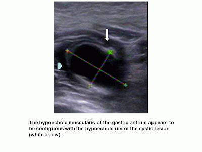
MSCT  without CE of abdomen detected  one mass with  intraluminal air and its wall was thickened more than 1cm which suggested inflammation like enteronecrosis.


This patient promptly was sent to BINH DAN HOSPITAL.and  abdomen x-ray for  check up  was done [see  photo].


Blood tests=   WBC rising 17K with  88% neutrophil.

Emergency operation performed as about peritonitis.due to perforation.
This mass is of  small intestine.which was looked like tumor or inflamation.


.

Microscopy reports that a cancer, But we have to wait for immunohistostainning to make sure that a malignant GIST or Carcinoid.

REFERENCE

Sunday 15 September 2013

CASE 209: A LUNG MASS, Dr PHAN THANH HẢI, MEDIC MEDICAL CENTER, HCMC, VIETNAM

Man 59yo in check-up lung x-ray detected a mass at left lung, asymptomatic, no history tuberculosis, non smocking patient.


Ultrasound of the lung showed that was a cystic avascular with well bordered mass, size of 4 cm.


MSCT CE said that cystic mass bilobar looked like a bronchogenic cyst.


Operation via endoscopic thoracotomy, and punction of this mass leaking out the milky fluid. Resection this tumor with macrospecimen picture.
Wait for pathology report, but the surgery report said it looked like a caseum cyst of tuberculosis.

REFERENCE:

Monday 9 September 2013

CASE 208: RETROPERITONEAL EXTRAUTERINE PREGNANCY, Dr PHAN THANH HẢI, MEDIC MEDICAL CENTER, HCMC, VIETNAM.

WOMAN  26yo UNDERWENT in vitro fertilization–embryo transfer (IVF–ET) WAS WAITING FOR THE RESULT.

BETA HCG  WAS RISING UP FROM 9K  TO 17K  BUT ULTRASOUND  CANNOT FIND OUT THE   INTRAUTERINE NIDATION.

AT THE RIGHT  OVARY SITE,  ULTRASOUND DETECTED ONE  MASS OF 2 cm WITHOUT BLOOD SUPPLY WHICH WAS  BIG AND  CYSTIC HEMORRHAGIC, NEAR RIGHT ILIAC VEIN.
.


FIRST LAPAROSCOPY DETECTED NOTHING.
ONE WEEK LATER   MRI WAS PERFORMED ALSO DETECTED  THIS MASS WHICH WAS  GROWING IN RETROPERITONEAL SPACE, NEAR  RIGHT ILIAC VEIN. Beta HCG AT THAT TIME WAS  UP TO  39K.


OPEN  SURGERY  REMOVED THIS  MASS, SUSPECTED EXTRAUTERINE PREGNANCY 
WAIT  FOR   PATHOLOGY REPORT  AND  FOLLOW UP THIS CASE  AS  EXTRAUTERINE PREGNANCY IN RETROPERITONEUM.

Tuesday 27 August 2013

CASE 207: GASTRIC DUPLICATION CYST, Dr PHAN THANH HẢI, Dr NGUYỄN THIỆN HÙNG, Dr TRẦN NGÂN CHÂU, MEDIC MEDICAL CENTER, HCMC, VIETNAM

A 26 year-old male patient  from Kien Giang province with chief complain: nausea and mild epigastric discomfort for 3 years, no vomiting, no fever. Family and his past medical history : nothing abnormal detectable. Physical examination: mild epigastric tenderness,  no mass in the epigastric area.


Undergoing of gastroendoscopy he was revealed a submucosa mass in the antrum which was confirmed  by CT; it was  a fluid-attenuation cystic mass in close  with the stomach wall but report of CT cannot rule out a heterotropic pancreas.


Ultrasound detected an 27x17mm intragastric cyst which adhered the greater curvature. The cyst wall had 2 layers: echogenic inner mucosal lining and hypoechoic outer rim which was contiguous with the muscular layer of the stomach. So we met a muscular rim sign of a  non-communicating GDC (gastric duplication cyst) in adult.


BD Hospital confirmed the non communicating GDC by filling defect on upper gastrointestinal series with barium meal and abdominal endoscopy exploration.


And  laparoscopy  was done to remove the GDC.


Wait for microscopic result.

Wednesday 21 August 2013

CASE 206: EPIGASTRIC MASS POST- PROSTATECTOMY for 5 YEARS, Dr PHAN THANH HẢI. MEDIC MEDICAL CENTER, HCMC, VIETNAM

Ultrasound check up  a 62 yo man , who underwent  prostatectomy for 5 years, detected one hypoechoic epigastric mass, size of 5cm in relation with great curvature of stomach. Color Doppler showed vascular supply from gastric artery.


Gastro-endoscopy detected no lesion inside stomach.

MSCT with CE showed this tumor was from gastric wall, and pediculated.


Blood test were normal all cancer markers: PSA, CEA , CA 19-9, CA 72-4.

Operation  laparotomy..showed this tumor is from  the  great curvature of stomach   with  long pedicule. Resection tumor see  macroscopy, wait for  microscopic report.


Saturday 17 August 2013

CASE 205: MEDIC RADIOLOGY CASE 9= Noncompacted Cardiomyopathy on MSCT 640, NGUYEN TUAN VU, NGUYEN THI KIM SANG, DUONG PHI SON, PHAN THANH HAI, MEDIC MEDICAL CENTER, HCMC , VIETNAM


HISTORY
Female patient , 33 yo, presented by severe heart failure for  1 year, previous diagnosis : dilated cardiomyopathy . Decreased S1. audible S3,  2/6 apical systolic murmur.  She was sent to MEDIC for cardiac MSCT to rule out Coronary artery disease.
EKG
Short PR, delta waves, QS in V2-V6, D1 aVL 


ECHOCARDIO+ TDI and 3 D Echocardiography
Decreased LV systolic function , LV diastolic dysfunction , LV diastolic dysfunction
Prominent trabeculae, spongiformed LV , Diagnostic criteria NC/C leyer > 2
Noncompacted cardiomyopathy Echocardiography: Apical 4 C view and Parasternal short axis view
Prominent trabeculations and spongiformed myocardium of LV 


MS CT 640               
Intertrabecular Recesses, Multiple Trabeculae , Predominant location at Apex, mid lateral, mid inferior . NC layer/ C layer > 2,2, Involving >2 segments, Sens. 100%, Spec. 95% (Tomography, volume 6, Issue 5, Sept.-Oct. 2012, pp 346-354)
MSCT 640: 3D Imaging =Trabeculated and spongiformed LV 


 Video clip from apex view


Summary
  Reported by Engberding and Benber in 1984 :Mutation in LDB3, genetic cardiomyopathy
  Myocardial sinusoids
  Severe heart failure, arrhythmias, thrombus formation, sudden death
  Diagnosis by  Echocardiography, MRI, MSCT
  Medical treatment ( ACEI, Betabloker, Aspirin, Anticoagulant ), ICD, heart transplant.
  Long term prognosis is unknown
  Value of cardiac MSCT in patient with heart failure.