Total Pageviews

Wednesday 2 January 2013

CASE 162: INTRAMUSCULAR MASS of RIGHT THIGH, Dr PHAN THANH HẢI, MEDIC MEDICAL CENTER, HCMC, VIETNAM


Woman 21 yo, painful sitting for 3 months at posterior site of the right thigh which irradiated to right foot.
Ultrasound 
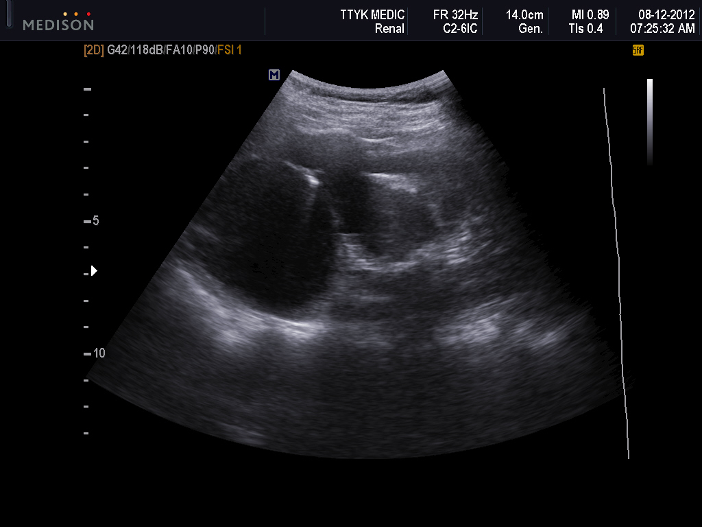
at the posterior of right thigh detected one intramuscular elliptical mass of 15cmX 4 cm, hypoechoic, well-bordered.

Image 1: longitudinal scan at posterior site of right thigh.

Image 2 : longitudinal section in CDI, this mass near deep femoral artery.

Image 3 : cross-sectional scan.

Image 4 : cross-sectional scan with linear 12 MHZ.

2 pictures of MSCT  without CE injection.


THE CORE BIOPSY REPORT OF THIS MASS IS NEUROMA OF SCIATIC NERVE.


DISCUSSION:  IT IS INTRA MUSCULAR TUMOR IN POSTERIOR SITE OF THE RIGHT THIGH,  HYPOECHOIC,  HYPOVASCULAR ,  FUSIFORM , ALONG THE SCIATIC NERVE TRAIL. THE CROSS-SECTION  OF THIS TUMOR BY HIGH FREQUENCY LINEAR PROBE IS TYPICAL  NERVE STRUCTURE.


 CROSS-SECTION  ANATOMY REFERENCE


Wednesday 26 December 2012

CASE 161: Vaginal Lymphangioma, Dr Tô Mai Xuân Hồng, Dept. of Ob-Gyn, University of Medicine and Pharmacology in HCM City


CASE 160: CAROTID ULTRASOUND, Dr PHAN THANH HẢI, MEDIC MEDICAL CENTER, HCMC, VIETNAM


Man  82 yo had been endarterectomy at right carotid artery  5 years ago.
Vascular ultrasound of carotid arteries for  check up.

Images  1, 2 : Right  carotid artery


Images  3, 4 : Left carotid artery

 
Do you see  any abnormalities on these pictures?  
 
 
The image of right carotid after endarterectomy is interesting by the wall ondulation like sea wave, but the flow is good.

On the left carotid , there is  big calcification plaque, and doppler artefact is no flow (see image pw )


Wednesday 19 December 2012

CASE 159: INTRAMUSCULAR MASS, Dr PHAN THANH HẢI, MEDIC MEDICAL CENTER, HCMC, VIETNAM


Man 28 yo, one year ago self detected one intramuscular mass of adductor muscle of the right thigh and pain with position sitting.
On ultrasound it is an intraadductor muscle ovoid mass,  size of 2x3cm, hypoechoic, posterior enhancement  and well bordered.

Doppler shows some intratumoral vessels (3  longitudinal section pictures with CDI).


REMOVED TUMOR, MACROVIEW.


Microscopic result is Schwannoma.
Reference

Sunday 16 December 2012

CASE 158: CYSTIC MASS NEARBY URINARY BLADDER, Dr PHAN THANH HẢI, MEDIC MEDICAL CENTER, HCMC, VIETNAM


Man 19 yo, onset acute abdominal pain, periodic crisis. Ultrasound emergency shows  one cystic mass with multilayer wall, with size of 5cm near urinary bladder. Intralumen of this cyst is one solid mass without vascular detection by Doppler (see 3 pictures US scan.)


MSCT abdomen is done, report this mass is one part of dilated ilium. What is this ?


Emergency operation with diagnosis of  small bowel torsion.


The surgeon reports that case of  intussusception by a small pediculate polyp.


Thursday 13 December 2012

CASE 157: THYROID CYST with COMET TAIL ARTIFACTS, Dr PHAN THANH HẢI, MEDIC MEDICAL CENTER, HCMC, VIETNAM


MAN 65 YO DETECTED HIMSELF RIGHT NECK TUMOR.

ULTRASOUND DISCLOSED THAT WAS A CYSTIC TUMOR  AT THE RIGHT LOBE THYROID WITH MANY COMET TAIL ARTIFACTS INSIDE THE CYST.

ASPIRATION THIS CYST REMOVED BROWN COLLOID FLUID.
CELL BLOCK REPORT WAS BENIGN CYST.


WHAT IS THE CAUSE OF COMET TAIL SIGN?
Reference:

Saturday 8 December 2012

CASE 156: RENAL CYST or HYDRONEPHROSIS, Dr PHAN THANH HẢI, MEDIC MEDICAL CENTER, HCMC, VIETNAM


WOMAN 49F PAIN AT LEFT SUBCOSTAL AREA.

 ULTRASOUND ABDOMEN DETECTED LEFT KIDNEY ABNORMAL WITH CYSTIC MASS AT UPER POLE.

WHAT IS YOUR DIAGNOSIS, RENAL CYST OR HYDRONEPHROSIS ? ( 2 IMAGES LONGITUDINAL  AND CROSS-SECTIONAL SCANS WITH B MODE GREYSCALE).

WITH B MODE IT IS VERY DIFFICULT  TO MAKE SURE DIAGNOSIS. WE USE PULSE DOPPLER, IT HAS  WAVEFORM PATTERN.


For  a clear diagnosis, we  use  CDI with  YINGYANG SIGN and  MSCT  UROLOGY which MAKE SURE THAT an AVM at the left kidney.


Summary= Black and white ultrasound  sometimes  makes mistake on diagnosis  of vascular diseases.


Reference