
Man 65 yo with abdomen distention (photo). For 40 years he
underwent a laparotomy in emergency by gunshot.

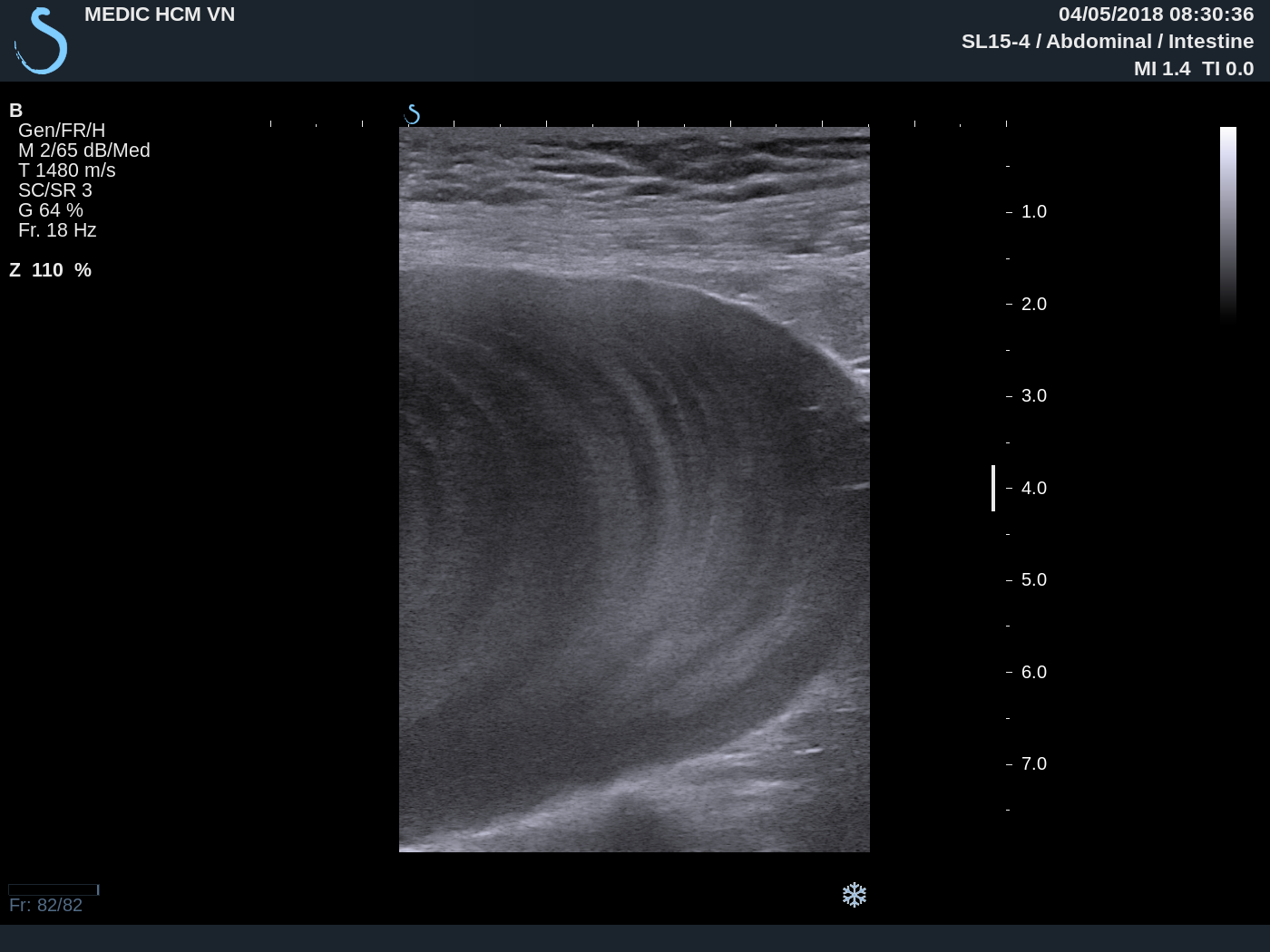
Ultrasound of abdomen detected at pelvis one round bordered
mass, size of 20cm. Its structure looked like cyst with many


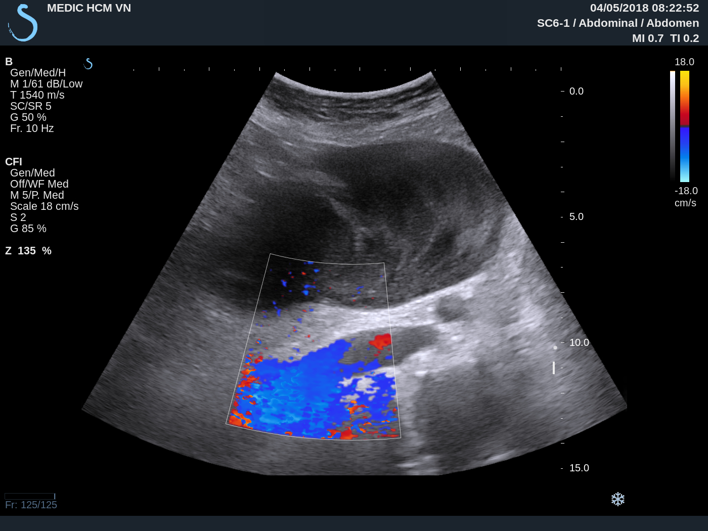
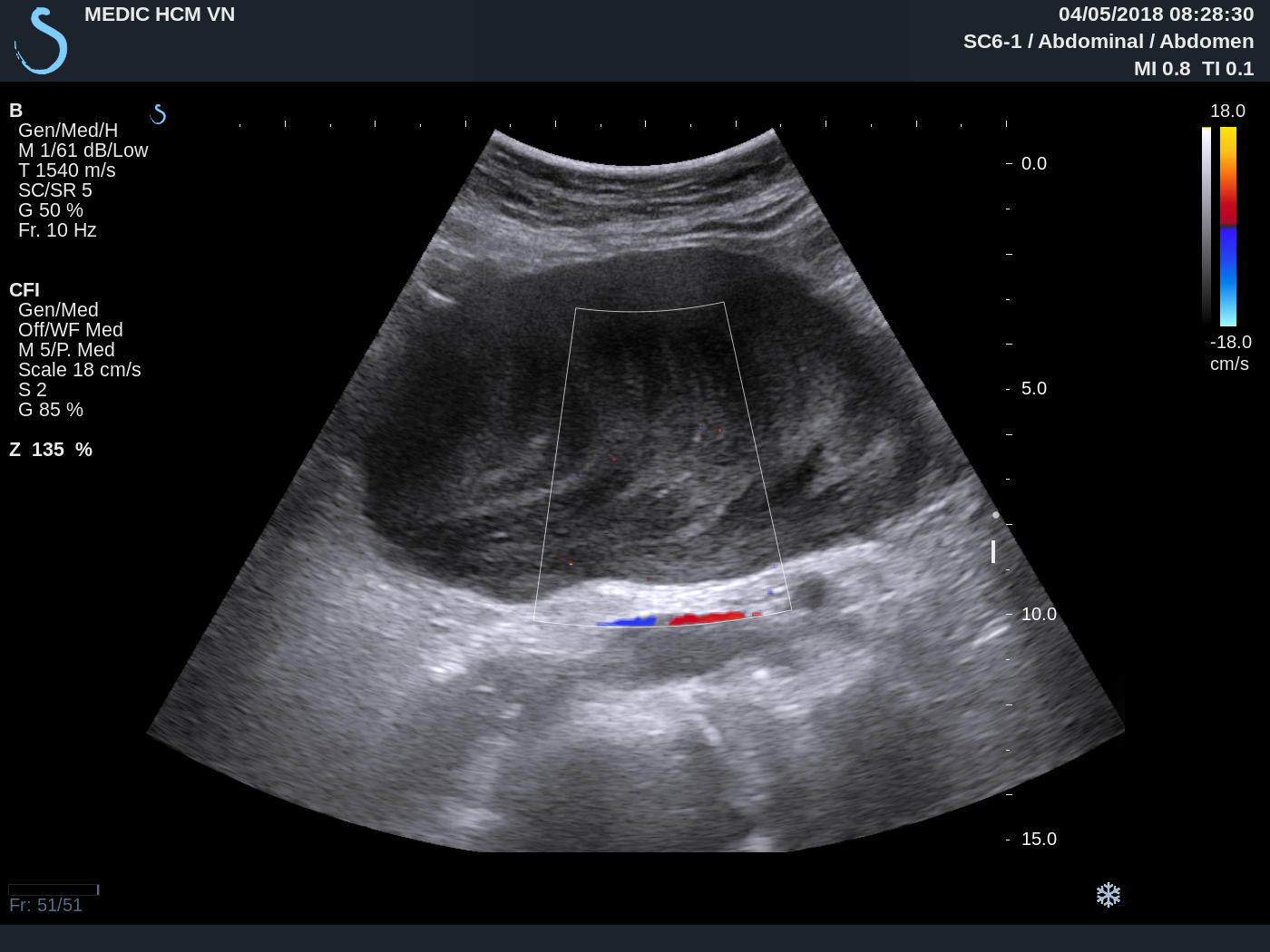
US 1: crossed- section at middle abdomen; US 2 : with
CDI, mass no vascular inside; US 3: longitudinal scan over
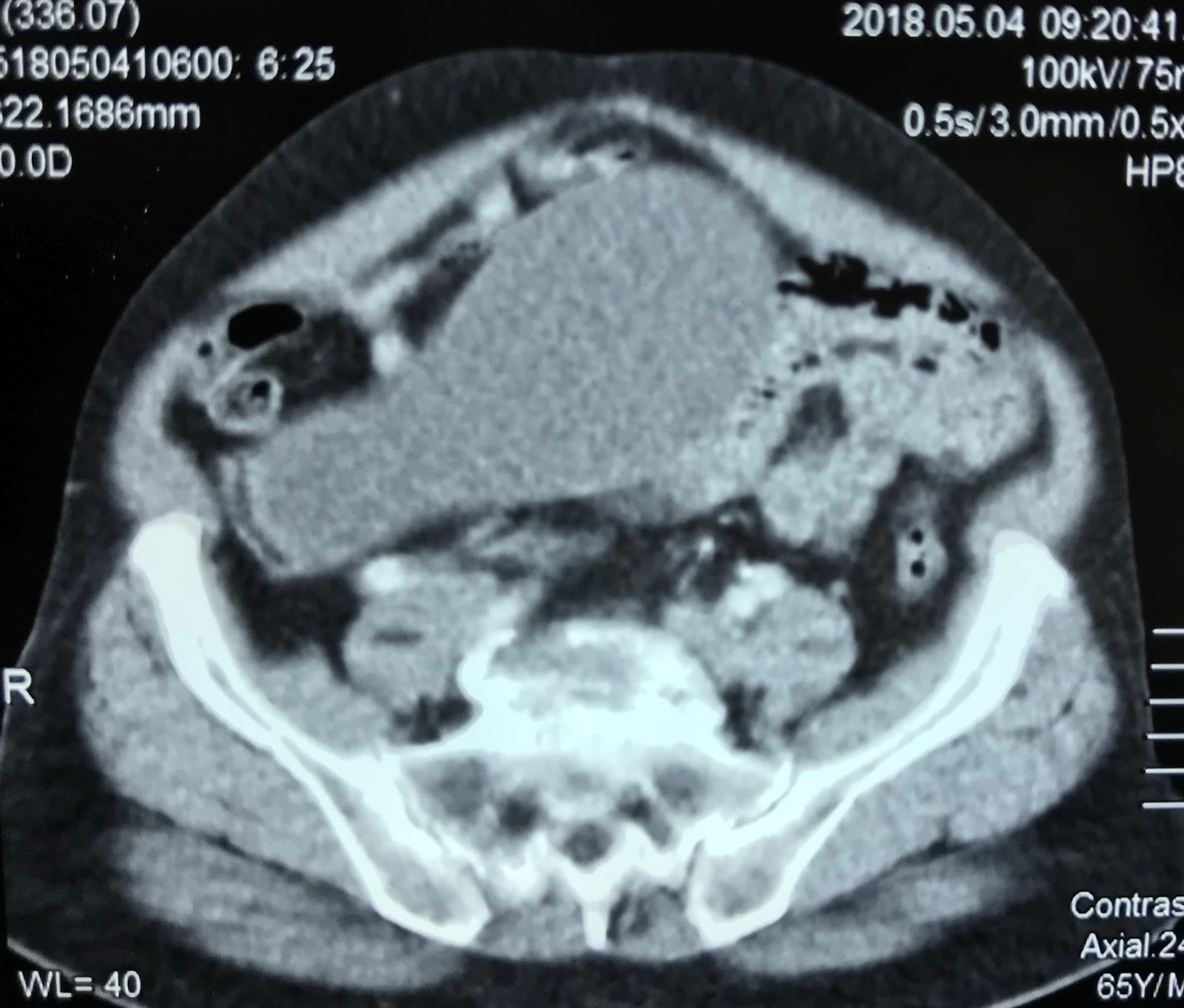
MSCT scan with
CE : CT 1: this mass is cystic formation from the coecum; CT 2 : frontal view.
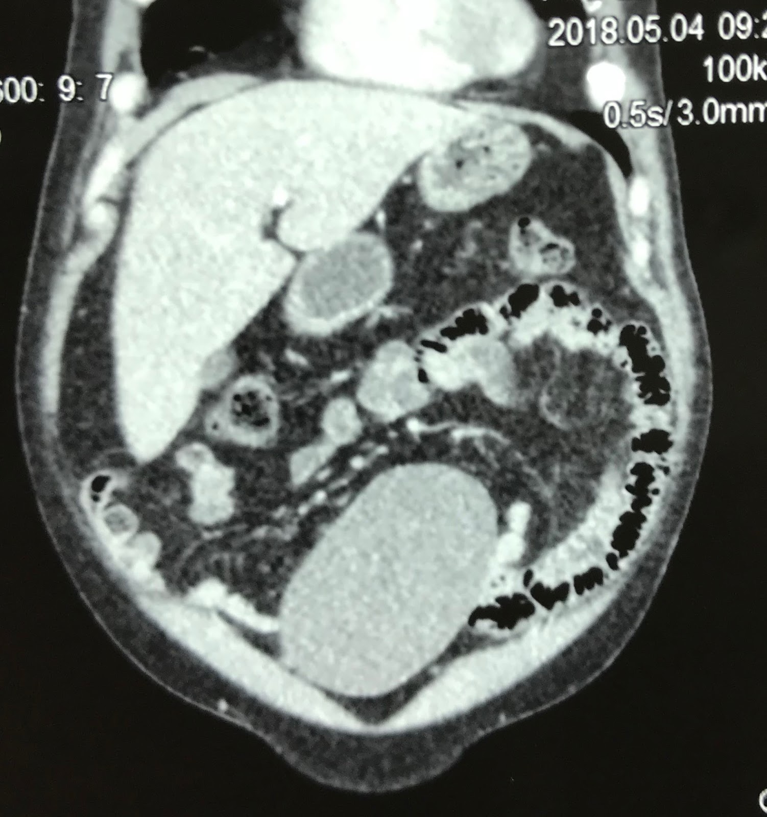
Appendicular mucocele
was made for diagnosing of the pelvic
mass. Operation removed one mass with mucus content from appendix.
DISCUSSION:
http://www.ytetunhantphcm.com.vn/vi/hoat-dong/khoa-hoc-dao-tao/82-ban-luan-ve-benh-u-nhay-ruot-thua-mucocele-of-the-appendix
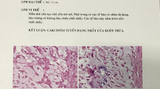
Microscopic report is mucineous cystadenocarcinoma.
REFERENCE:
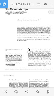
https://onlinelibrary.wiley.com/doi/pdf/10.7863/jum.2004.23.1.117
.DISCUSSION:
http://www.ytetunhantphcm.com.vn/vi/hoat-dong/khoa-hoc-dao-tao/82-ban-luan-ve-benh-u-nhay-ruot-thua-mucocele-of-the-appendix
Microscopic report is mucineous cystadenocarcinoma.

REFERENCE:

https://onlinelibrary.wiley.com/doi/pdf/10.7863/jum.2004.23.1.117
No comments :
Post a Comment