
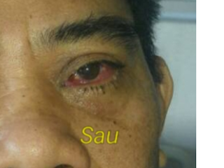
Man 52 yo with cough and pain at left
eye, protrusion the orbis and edema the cornea for one week (photo).

MRI of the eye and or brain.


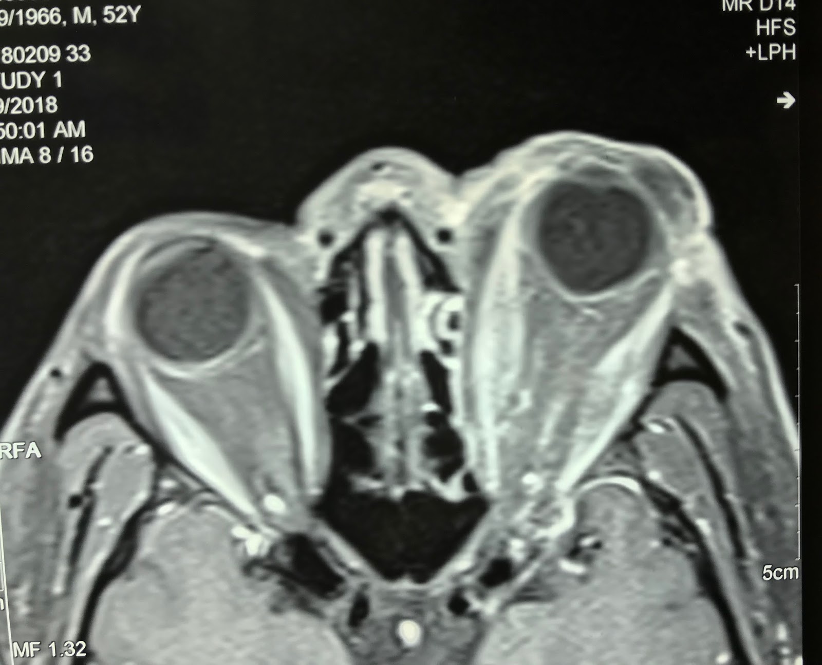
MRI 1= frontal view of the left exopthalmic
eye.
MRI 2= frontal section of the left orbis. edema
of the intra orbis muscles,
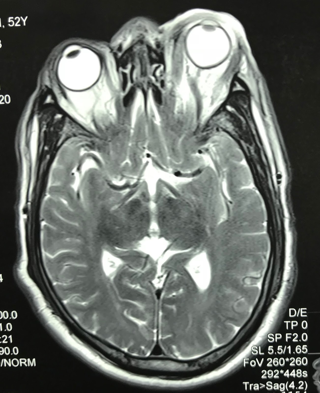
MRI 3= crossed section of left
orbis : left superior opthalmic vein dilated.
MRI 4= the muscles in orbis are
edema and cavernous sinus is not abnormal.
For make sure diagnostic DSA was done that detected A-V
fistula at cavernous sinus.
DSA with dilated opthalmic vein= DSA1
putting of the coil, DSA 2 after treatment. Coil embolisation
is spectacular reduction clinical sign (photo 2).


The left eye returns near normal 24 hrs after treatment.
Conclusion: Basis clinical signs of MRI and DSA can make diagnosis and spectacular treatment. success.

Conclusion: Basis clinical signs of MRI and DSA can make diagnosis and spectacular treatment. success.
Reference: Anatomy of eye circulation.

No comments :
Post a Comment