
Woman 59yo detected one mass at right buttock that
is in slow growth and no painfull.
Ultrasound scan
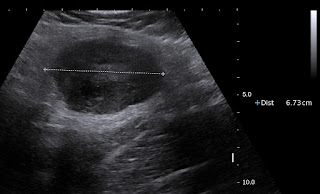
US 1= longitudinal scan of this mass with size 6.7 cm,
ovoid, hypoechoic in gluteus maximus muscle near sciatic
nerve like a size of a mango.

US 2 = crossed section of this tumor is well bordered
and hypovascular pattern.


US 3 = elasoscanning of this mass is inhomogeneous
structure.

MRI scan


MRI 1 crossed section this mass is
well bordered in a muscle
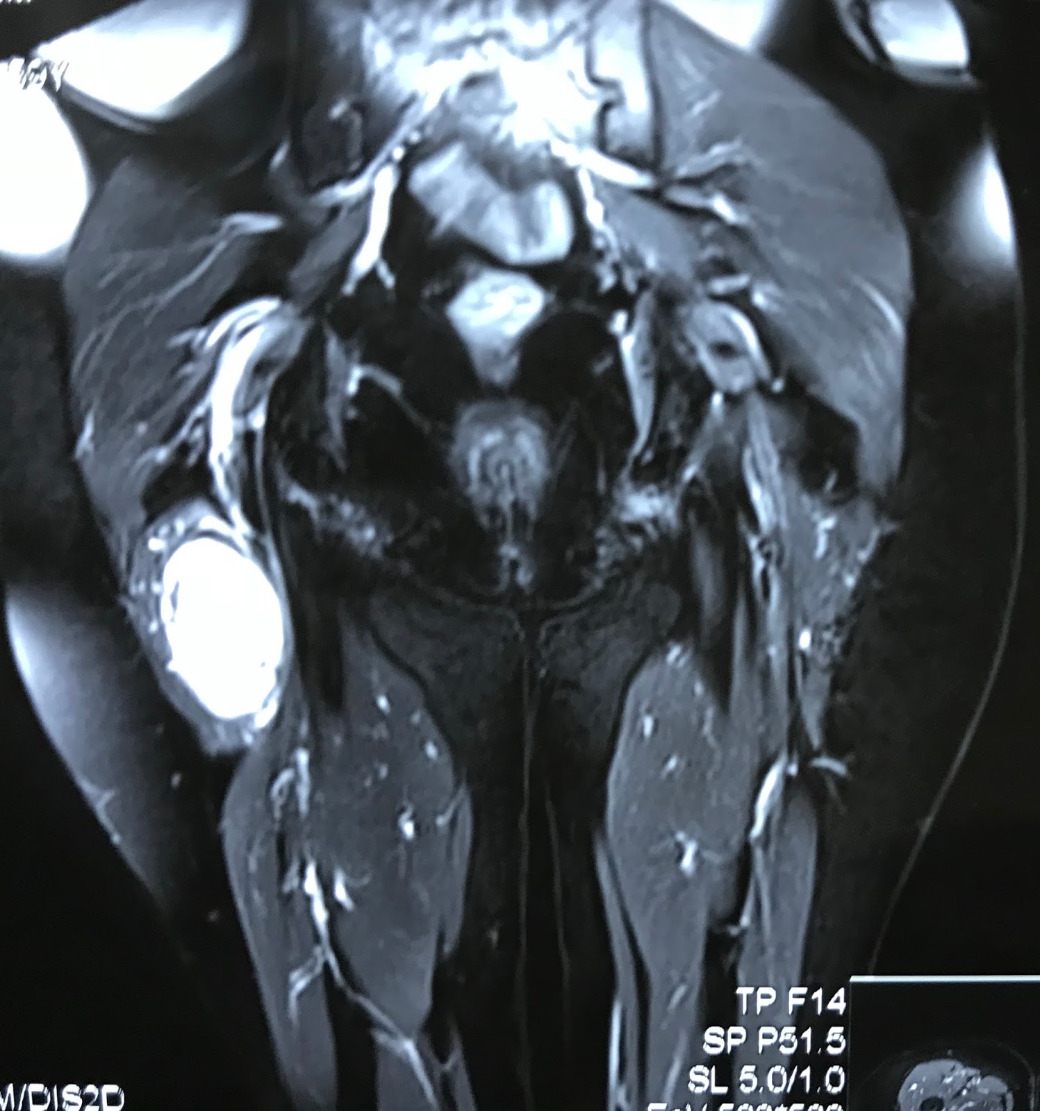
MRI 2 frontal view this mas is bordered of right sciatic
nerve.
MRI 3 relation of this mass and right sciatic nerve.


Core biopsy is done and histology report
is neuroma.
No comments :
Post a Comment