Man 67 y o,
emergency operation by acute necrosis of gallbladder by stone one week ago, still
pain at Murphy area. WBC = 12k with neutro 90%, CRP=
100ng/mL.
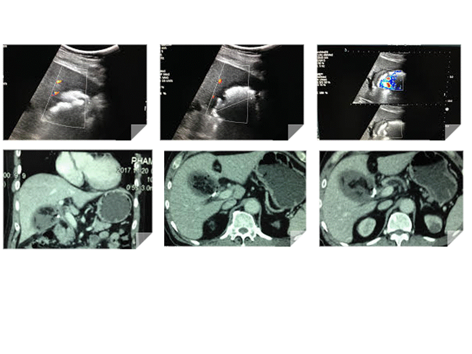
Abdomen ultrasound
detected one mass # 5 cm at the bed of gallbladder. Mass has got fluid content and white structures inside with very
strong shadowing and air in formatting an abscess.
No dilatation
of the biliary system.
US 1:
subhepatic abscess with strong shadowing in abscess.

US 2: umbrella sign of the
shadowing.
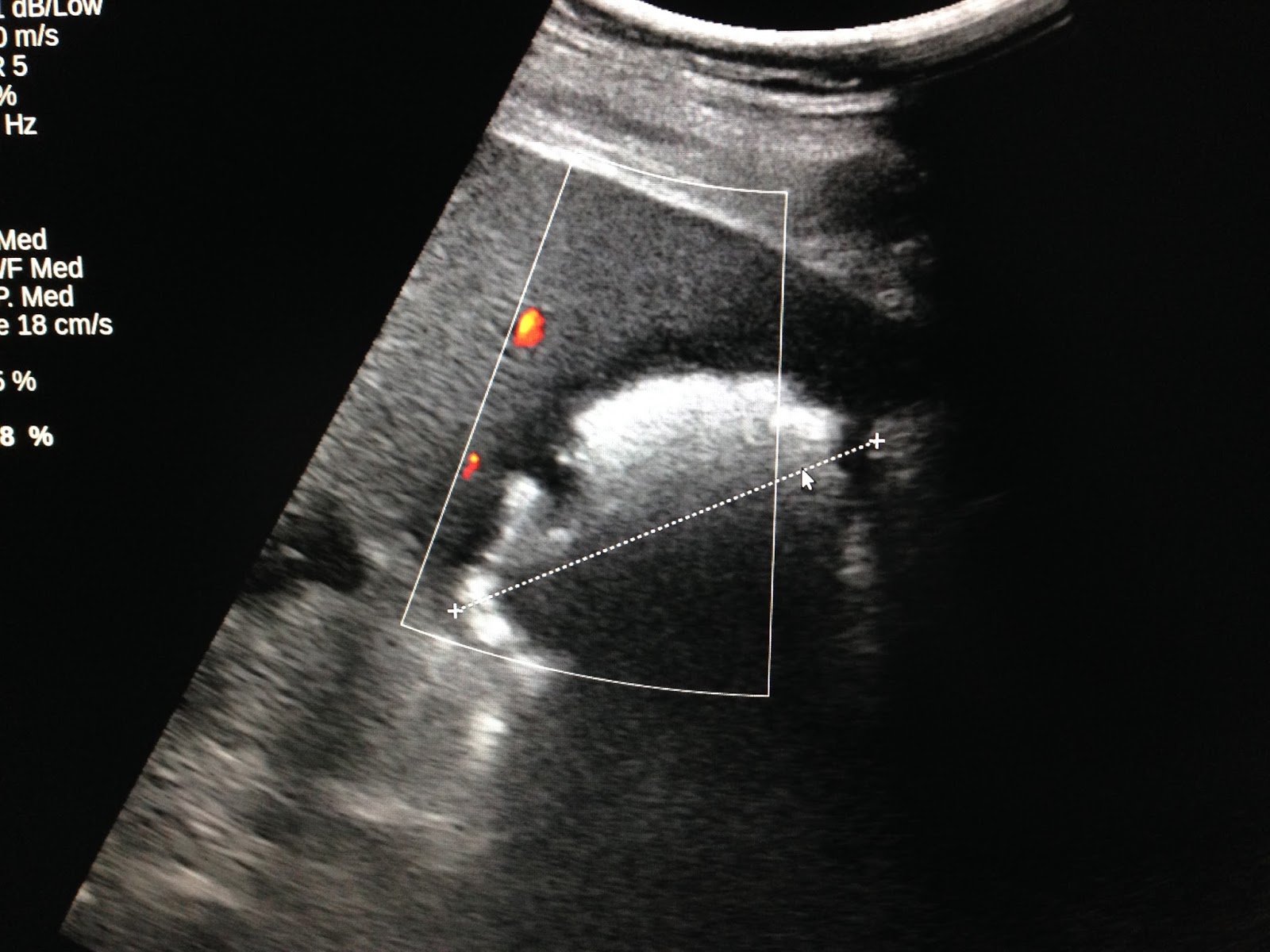
US 3:
elatoscan shows this structure is very hard.

MSCT:
CT 1: abscess with air and fluid filling at the bed of gall
bladder which had been removed of GB.
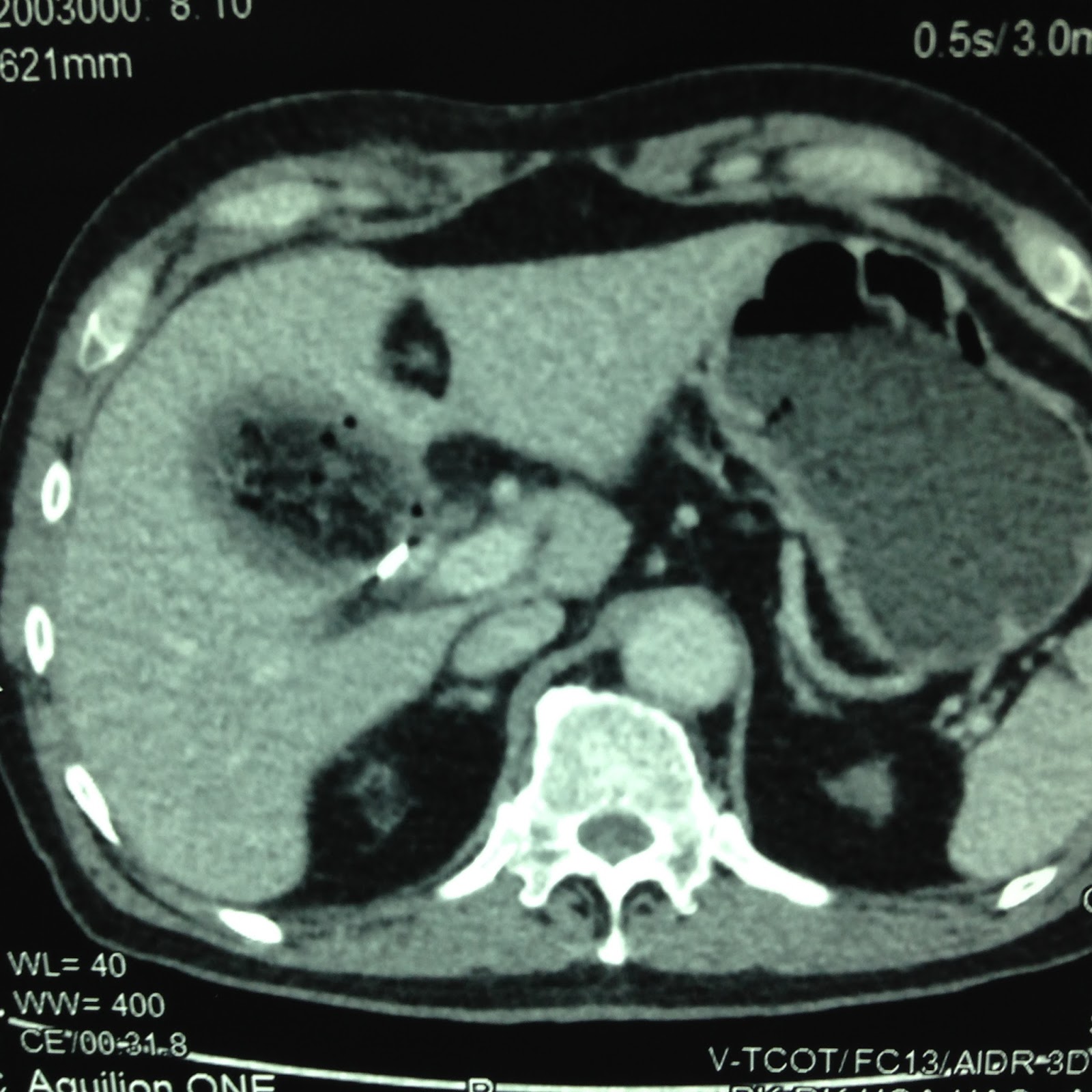
CT 2:
crossed-section view of this abscess:
inhomogenous structure and air

CT 3: frontal view of the
abscess.

Radiologist
reported textilloma in suspection.
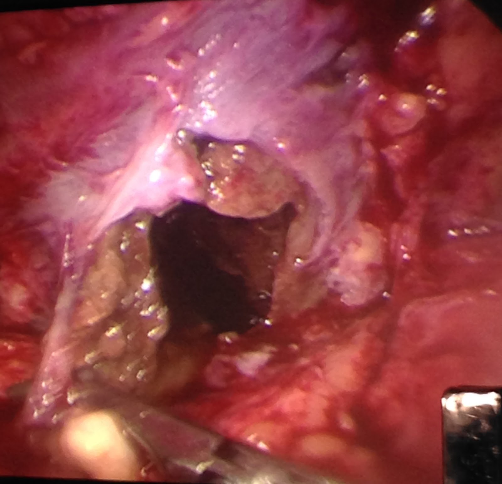
Laparoendoscopy detected an abscess in liver at the bed of gallbladder necrosis and no textilloma.
No comments :
Post a Comment