
Man 36yo with right testis tense.
Ultrasound scanning of right scrotum detected big testis focal
lesion, round, size of 3cm
US 1 color doppler not hypervascular, well bordered.

US 2 CDI: hypovascular tumor.

US 3 elastoscan of this tumor is inhomogeneous with some
parts very hard.

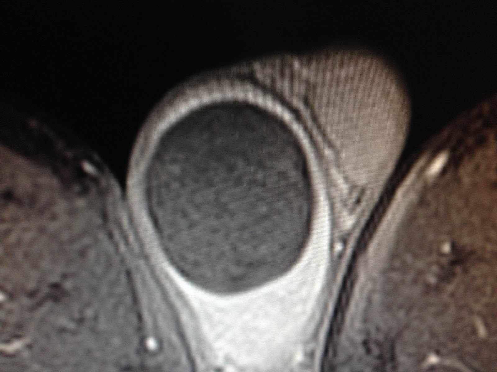
MRI with gado: MRI 1, 2, 3: this tumor very low gado enhanced.


Radiologist suggested epidermoid cyst.
Blood tests = normal AFP and HCG.
Operation resection of right testis (see macro 1, 2).

Microscopic report is epidermoid cyst.

No comments :
Post a Comment