
Woman 57 yo
detected on mass below the xyphoid process at middle line.
Ultrasound scan of the
epigastric region of abdomen with curve probe 3.5 MHz.
US1: longitidinal scan detected the
defected abdomen wall and the mass is connected into
abdomen.

US 2: crossed section.of.this
mass.

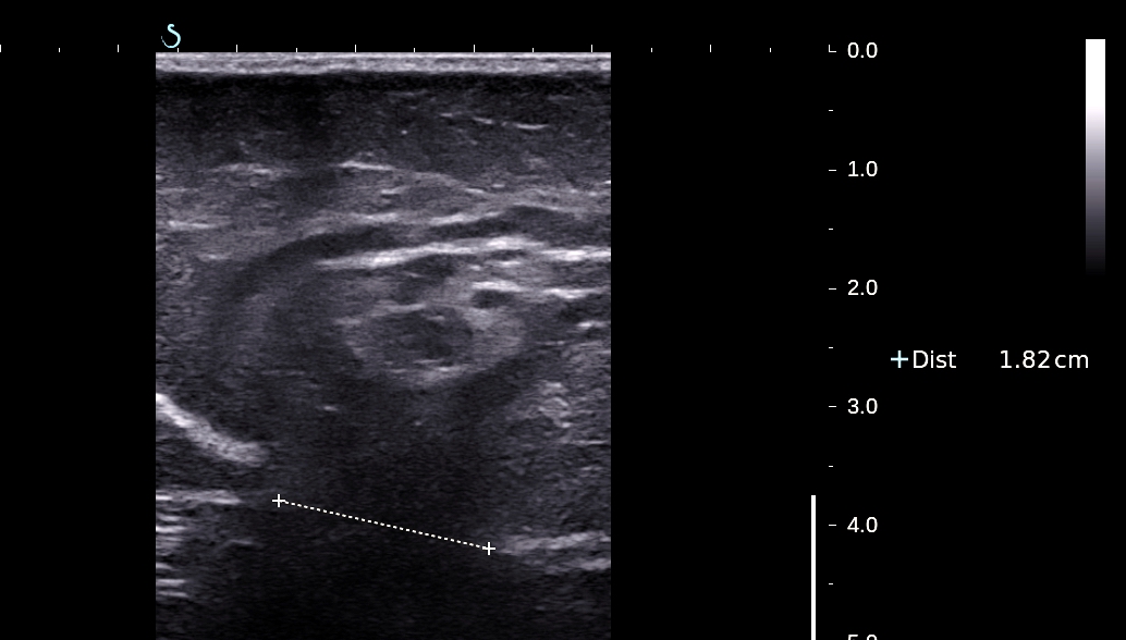
US 3 scanning of
this mass with linear probe 10 MHz. Diameter of the orifice #
1.5cm. And fatty tissue is
pulled out the abdomen by this orifice.
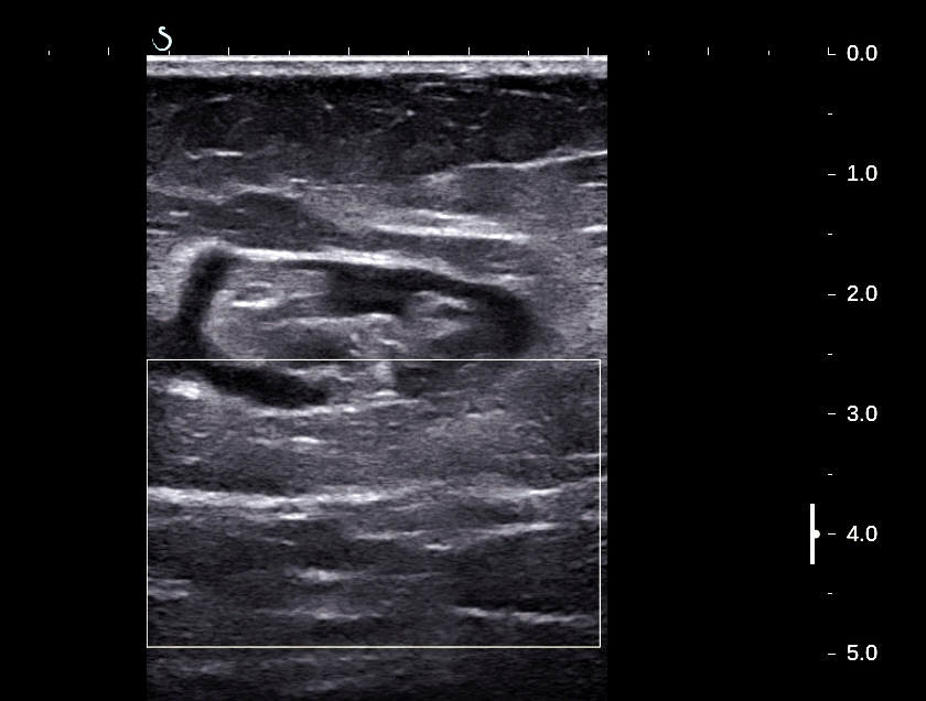
US 4 : crossed
section of this mass.
Sonologist diagnosed
epigastric hernia.
Operation showed this
mass is built by fatty tissue and epiploid of transverse
colon.

Picture reference.

No comments :
Post a Comment