Women
30yo,
general check- up .
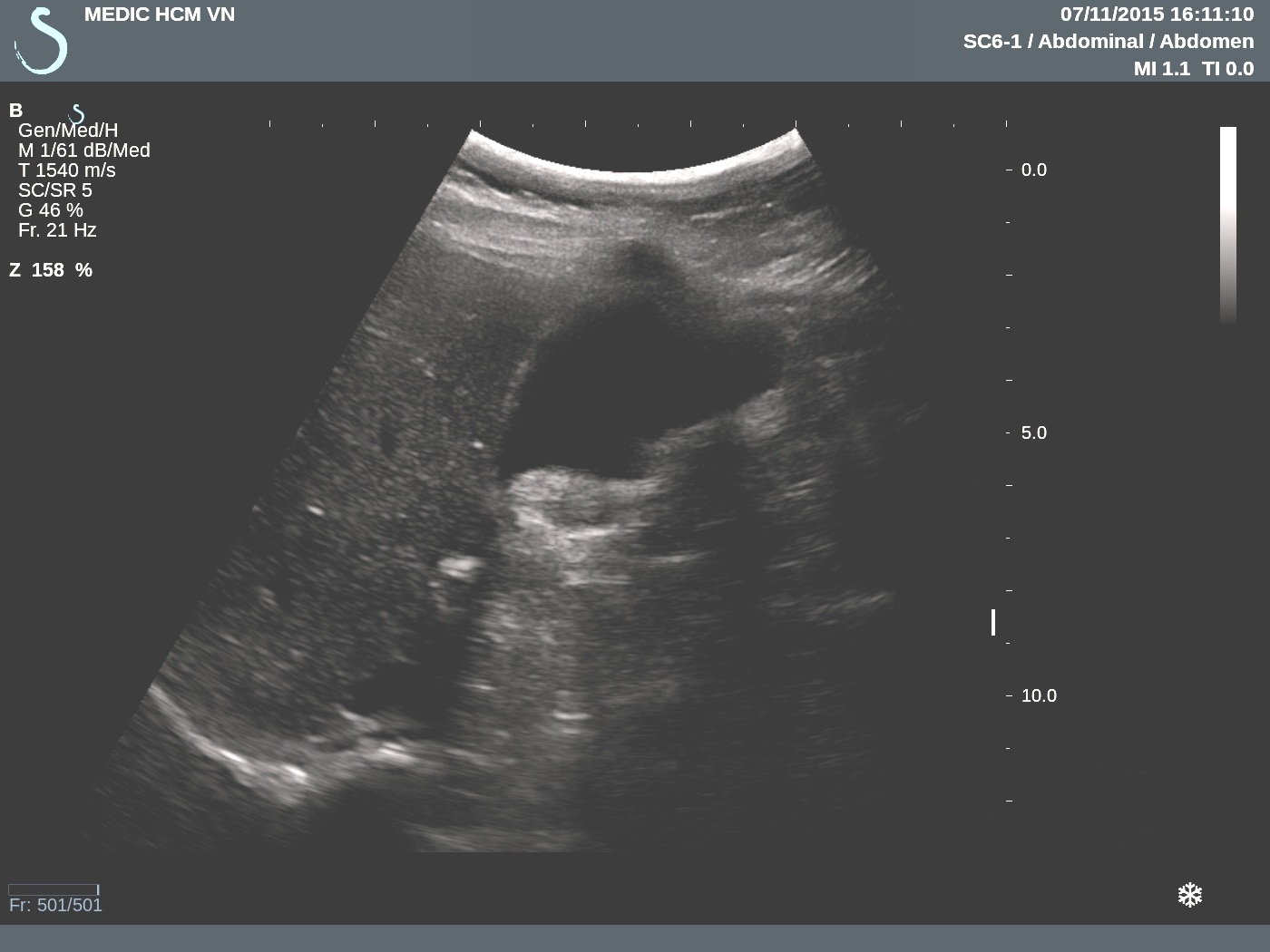
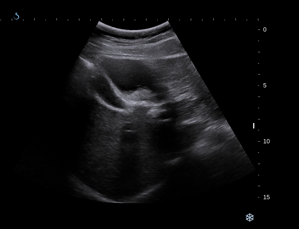
Ultrasound detected a
tumor on border of liver near gallblader which deplaces left
gastric curvature and is from retroperitoneal
space. Its
structure are solid and cystic parts, size arround 10cm ( see ultrasound us1.. cystic part
tumor in border liver; us 2..near gallblader; us 3..long scan left lobe liver and
tumor.). Sonologist cannot diagnose this tumor from lesser omentum.

MSCT with CE
of this tumor is mixed structure, cystic,
fatty, and calcification [ CT1..section, CT 2
frontal section , CT3 sagital ). Suggession from radiologist is teratoma tumor
or lipoma necrosis.


MRI
with gado ( MRI 1..struture is more fat tissue., MRI 2..with
fat suppression , MRI 3 frontal
view). Radiologist says
teratoma
of retroperitoneum, in
lesser omentum area.


Blood
test of
all cancer markers are
normal.
Laparo-operation= 

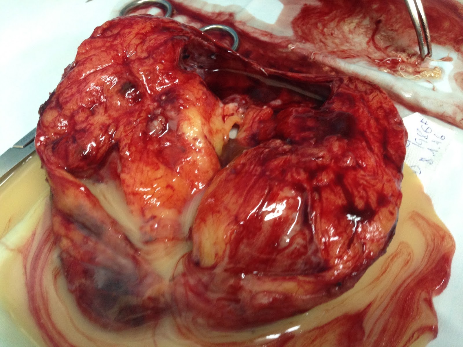
picture 1( retrogastric tumor well bordered)
picture 2macro
picture macro 3, opened specimen, solid and cystic tumorand fluid inside like milk)
Microscopic report of this tumor is teratoma maturation.

No comments :
Post a Comment