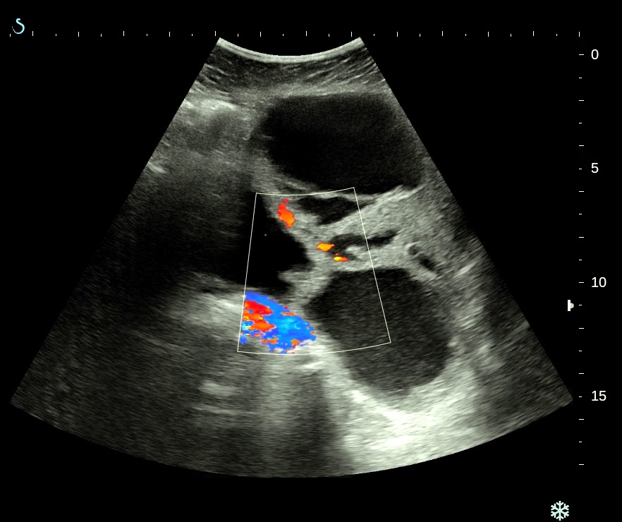
Woman 50 yo, vaginal bleeding.
Pelvis ultrasound detected one mass cystic, multiloculated
with septation thickening and solid part. No ascites (see
5 US pictures).


MRI with gado reported with enhanced CE suspected ovary cancer.


Blood test : CA-125 rising 125 U/mL
Pre operative diagnosis is ovary cancer stage II B. Microscopic specimen report is serous cystadenocarcinoma.

Discussion: With 3 modalities for diagnosing this case ULTRASOUND, MRI and BLOOD TEST MARKER, what is the excellent value?.
No comments :
Post a Comment