Woman 75 yo vomiting, endoscopy detected an extragastric fundus tumor (see pictures).

Ultrasound of abdomen
showed one hypoechoic mass with size of 4 cm, well-bordered at the hilus of spleen ( see 2 ultrasound pictures).


MSCT with CE found out this mass bending the wall of
great curvature of stomach, very slow CE enhancement (see 3 CT pictures).


Blood tests of all markers are normal.
What is your suggestion of diagnosis?
Open surgery removed the tumor easily. It grew from gastric fundus wall, its structure was hard.


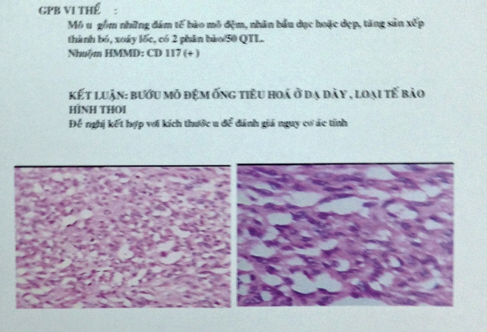
MICROSCOPIC REPORT WITH IMMUNOHISTO CHEMISTRY is GIST OF STOMACH WALL.
REFERENCE:


MICROSCOPIC REPORT WITH IMMUNOHISTO CHEMISTRY is GIST OF STOMACH WALL.


No comments :
Post a Comment